


#ExpertRead | Colorectal Surgery
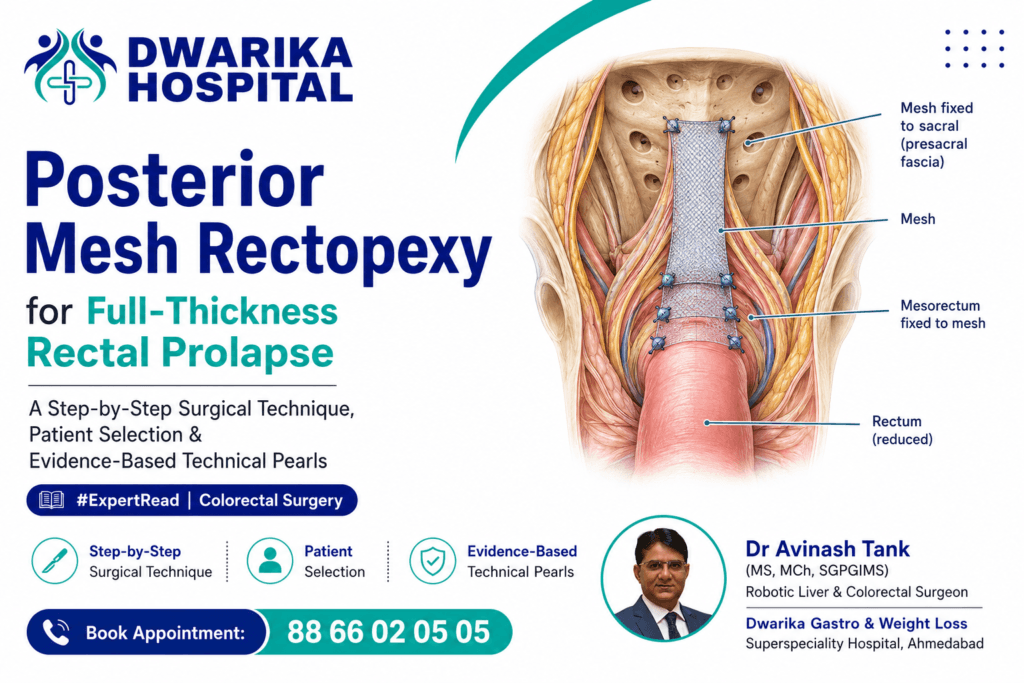
Posterior mesh rectopexy—classically described as the Wells procedure—is an abdominal rectopexy in which the rectum is mobilized posteriorly and fixed to the sacrum using a mesh. Although ventral mesh rectopexy has become increasingly popular, posterior mesh rectopexy remains an important option, particularly in centres with extensive experience with the technique.
The modern evidence does not justify describing one rectopexy as universally superior. Patient anatomy, constipation/incontinence phenotype, previous surgery, pelvic floor dysfunction, surgeon expertise and mesh-related considerations should influence the operation chosen. The 2017 ASCRS guideline specifically noted insufficient evidence to establish superiority of posterior versus anterior repairs. (fascrs.org)
1. What is posterior mesh rectopexy?
The fundamental principle is:
Mobilize the prolapsed rectum → restore it to its anatomical position → create durable fixation to the presacral fascia/sacrum.
Unlike ventral mesh rectopexy (VMR), where dissection is predominantly anterior and the mesh is attached along the anterior rectal wall, posterior mesh rectopexy involves posterior mobilization and posterior mesh fixation.
The classical Wells operation uses a mesh positioned behind the rectum and fixed to the sacral/presacral region.
Modern laparoscopic descriptions have modified the original operation substantially, particularly with regard to mesh size, fixation and peritoneal coverage.
2. When should posterior mesh rectopexy be considered?
The typical candidate is a patient with:
- Full-thickness external rectal prolapse
- Acceptable operative fitness for abdominal surgery
- Significant prolapse-related symptoms
- Need for a durable abdominal repair
- Anatomy suitable for laparoscopic/robotic rectopexy
- No contraindication to mesh implantation
The operation should not be selected solely because the prolapse is large.
The functional phenotype is equally important.
Before surgery, document:
Prolapse
- External versus internal prolapse
- Length of prolapse
- Solitary versus multicompartment pelvic organ prolapse
- Previous prolapse surgery
Bowel function
- Constipation
- Obstructed defecation
- Excessive straining
- Incomplete evacuation
- Fecal urgency
- Fecal incontinence
Anatomy
- Colon redundancy
- Rectosigmoid configuration
- Pelvic floor descent
- Enterocele
- Rectocele
- Uterine/vaginal prolapse where relevant
This is important because rectopexy is not simply a mechanical operation—it can change bowel function.
3. The operative concept
A useful mental model is:
REDUCE → MOBILIZE → POSITION → FIX → COVER
Step 1 — Establish laparoscopic access
A standard multiport laparoscopic approach can be used.
The patient is generally positioned in:
Modified lithotomy + Trendelenburg
The small bowel is displaced cranially to expose the pelvis.
A 2011 multimedia technical report described a four-port laparoscopic approach for posterior mesh rectopexy. (pubmed.ncbi.nlm.nih.gov)
4. Identify the rectosigmoid and sacral promontory
The first major landmarks are:
- Sigmoid colon
- Rectosigmoid junction
- Sacral promontory
- Right pelvic sidewall
- Hypogastric nerves
Technical principle
Do not begin deep pelvic dissection before identifying the autonomic nerves and the correct avascular planes.
This is particularly important because excessive lateral/posterolateral dissection can damage the sympathetic and parasympathetic pelvic nerves.
5. Posterior rectal mobilization
The rectum is mobilized posteriorly.
The dissection proceeds in the plane between:
mesorectum ↔ presacral fascia
and is carried down toward the pelvic floor.
The classical laparoscopic description specifically recommends posterior dissection to the level of the pelvic floor. (pubmed.ncbi.nlm.nih.gov)
The critical objective
Mobilization must be adequate to permit reduction of the prolapse without tension, but unnecessary lateral dissection should be avoided.
This is one of the most important technical balances in rectopexy.
6. How far should posterior dissection go?
A practical endpoint is:
Pelvic floor / levator level
The surgeon should be able to mobilize the rectum sufficiently that:
- the prolapse is completely reduced,
- the rectum lies without tension,
- fixation does not pull the rectum excessively upward,
- and the mesh can lie smoothly against the sacrum.
Over-dissection is not automatically better.
Excessive mobilization can contribute to:
- autonomic nerve injury,
- postoperative constipation,
- pelvic dysfunction.
7. Anterior mobilization—how much is necessary?
This is an important distinction between posterior mesh rectopexy and ventral mesh rectopexy.
In the posterior mesh technique, the primary fixation is posterior.
However, the original laparoscopic technique described by Zmora et al. included anterior mobilization to the pelvic floor before placement of the posterior mesh. (pubmed.ncbi.nlm.nih.gov)
Modern surgeons may modify the extent of anterior dissection depending on:
- associated rectocele,
- enterocele,
- intussusception,
- pelvic organ prolapse,
- previous surgery,
- functional symptoms.
Therefore, “posterior mesh rectopexy” should not be interpreted as a single universally standardized dissection technique.
8. Preparation of the sacral fixation point
The mesh is positioned vertically along the sacrum.
In the classic laparoscopic multimedia technique:
- approximately 5 × 2 cm mesh was introduced,
- positioned vertically on the sacrum,
- extending from around the sacral promontory downward,
- and fixed using endoscopic tackers. (pubmed.ncbi.nlm.nih.gov)
Important anatomical warning
Fixation should be performed:
below the promontory and close to the midline
to reduce the risk of injury to the hypogastric nerves. (pubmed.ncbi.nlm.nih.gov)
9. Mesh fixation to the rectum
Once the mesh is securely attached to the sacral region, the rectum is brought against it.
The mesorectum is then fixed to the mesh.
The original laparoscopic technique described four-point fixation with absorbable sutures. (pubmed.ncbi.nlm.nih.gov)
This creates:
Sacrum → mesh → mesorectum → rectum
rather than attempting to place multiple difficult sutures directly into the presacral fascia.
One proposed technical advantage is that suturing the mesorectum to the mesh is easier than placing sutures directly into the presacral fascia using laparoscopic instruments. (pubmed.ncbi.nlm.nih.gov)
10. The mesh should not strangulate the rectum
This is a critical technical pearl.
The mesh should provide:
Support—not constriction.
Avoid:
- excessive tightening,
- excessive upward traction,
- focal pressure on the rectal wall,
- twisting of the mesorectum.
Continue Reading — Free
Get the complete expert guide from Dr. Avinash Tank.
Free, always. No payment, no spam.
Continue Reading — Free
Get the complete expert guide from Dr. Avinash Tank.
Free, always. No payment, no spam — just the complete guide sent straight to your screen.
The rectum should remain anatomically positioned while preserving physiological compliance.
11. Peritoneal coverage
After mesh fixation, the peritoneum should be closed over the mesh where appropriate.
The objective is to:
- isolate the mesh from the abdominal cavity,
- reduce bowel contact,
- reduce adhesion formation,
- restore the peritoneal surface.
The reported Wells-type technique in the literature describes careful peritoneal coverage of the mesh. (pubmed.ncbi.nlm.nih.gov)
12. What are the most important technical danger zones?
⚠️ 1. Hypogastric nerves
The sacral fixation zone is close to the autonomic nerves.
Rule: Stay close to the midline when appropriate and avoid deep lateral fixation.
The technical report specifically emphasizes positioning fixation below the promontory and adjacent to the midline to avoid hypogastric nerve injury. (pubmed.ncbi.nlm.nih.gov)
⚠️ 2. Presacral bleeding
The presacral plane can produce troublesome bleeding.
Prevention is preferable to treatment:
Correct plane + controlled dissection + careful fixation.
⚠️ 3. Ureter
Particularly important during lateral pelvic dissection and in patients with previous pelvic surgery.
⚠️ 4. Rectal wall
Avoid incorporating the rectal lumen or causing excessive focal pressure.
⚠️ 5. Mesh-related complications
Mesh erosion, infection, fistulation and chronic pelvic pain remain important considerations whenever synthetic mesh is used.
13. What does Level-1 evidence actually tell us?
This is where an important distinction must be made.
There are excellent randomized trials comparing different rectopexy strategies, but there is relatively little high-quality randomized evidence specifically testing posterior mesh rectopexy itself.
Therefore, one should not overstate the evidence.
Randomized evidence
A double-blind randomized trial from Denmark compared:
Laparoscopic ventral mesh rectopexy vs laparoscopic posterior sutured rectopexy
in 75 patients with full-thickness rectal prolapse.
At 12 months, there was no significant difference in improvement of obstructed defecation symptoms between the two procedures. (pubmed.ncbi.nlm.nih.gov)
At six-year follow-up, the ventral mesh group had better constipation-related quality-of-life and symptom scores, but this was a relatively small single-centre randomized cohort and does not establish universal superiority. (pubmed.ncbi.nlm.nih.gov)
14. What does the broader comparative evidence show?
A 2021 meta-analysis of five comparative studies involving 307 patients found that laparoscopic mesh rectopexy had a lower recurrence rate than posterior sutured rectopexy:
OR 0.28; P = 0.009
but required longer operative time.
Importantly, there were no significant differences in constipation or incontinence scores. (pubmed.ncbi.nlm.nih.gov)
This suggests an important practical message:
Mesh may improve mechanical durability, but the functional consequences are not automatically better.
15. Network meta-analysis: an interesting signal for posterior mesh rectopexy
A 2023 network meta-analysis included 9 randomized clinical trials involving 728 patients.
Posterior mesh rectopexy ranked highly for recurrence outcomes and had lower estimated recurrence odds compared with several other operations, including:
- Altemeier
- Delorme
- resection rectopexy
- sponge rectopexy
- sutured rectopexy.
However, posterior mesh rectopexy was not significantly different from ventral mesh rectopexy for recurrence.
There were also no significant differences between procedures in complications, operating time or improvement in fecal incontinence. (pubmed.ncbi.nlm.nih.gov)
Important interpretation
This is powerful comparative evidence—but it is a network meta-analysis, not a large direct randomized trial of posterior mesh rectopexy versus every competing operation.
Therefore:
High-level evidence ≠ evidence that posterior mesh is universally the best operation.
16. The constipation question
This is perhaps the most clinically important issue.
Rectal prolapse patients may have:
Phenotype A
Prolapse + fecal incontinence
or
Phenotype B
Prolapse + constipation/ODS
or
Phenotype C
Prolapse + both
or
Phenotype D
Prolapse without major functional symptoms
The operation should be tailored accordingly.
Mesh rectopexy can correct the anatomical prolapse, but constipation is multifactorial.
Potential contributors include:
- slow colonic transit,
- pelvic floor dyssynergia,
- rectal hyposensation,
- excessive rectal fixation,
- internal intussusception,
- rectocele,
- redundant sigmoid colon.
Therefore:
Do not promise constipation relief simply because the prolapse will be corrected.
17. What about fecal incontinence?
Successful rectal prolapse repair frequently improves continence because prolapse itself can impair:
- anal sphincter function,
- rectal sensation,
- continence mechanics.
The 2023 network meta-analysis found no statistically significant differences among the evaluated operations in improvement of fecal incontinence. (pubmed.ncbi.nlm.nih.gov)
A 2024 systematic review specifically examining men reported substantial improvement in fecal incontinence following posterior mesh rectopexy, although this evidence is primarily observational and heterogeneous. (pubmed.ncbi.nlm.nih.gov)
18. Posterior mesh versus posterior suture rectopexy
| Feature | Posterior mesh | Posterior suture |
|---|---|---|
| Rectal mobilization | Posterior | Posterior |
| Mesh | Yes | No |
| Mechanical fixation | Mesh + fixation | Sutures |
| Recurrence | May be lower | Potentially higher |
| Operative complexity | Higher | Lower |
| Mesh complications | Possible | None |
| Constipation advantage | Not clearly established | Not clearly established |
| Evidence | Moderate comparative evidence, limited direct RCTs | More established randomized comparisons |
| Surgeon experience | Critical | Critical |
A 2024 meta-analysis similarly found lower recurrence with mesh rectopexy compared with suture rectopexy, but longer operative time and no significant difference in constipation improvement. (pubmed.ncbi.nlm.nih.gov)
19. Posterior mesh versus ventral mesh rectopexy
This is a much more interesting debate.
Posterior mesh
Advantages
- Strong posterior fixation
- Circumferential/posterior mobilization
- Familiar technique in many colorectal units
- Potentially excellent recurrence outcomes
Disadvantages
- More posterior/lateral dissection
- Greater concern regarding autonomic nerves
- Constipation concerns
- Mesh-related complications
Ventral mesh
Advantages
- Avoids extensive posterior rectal mobilization
- Preserves lateral ligaments
- Particularly attractive in selected patients with anterior compartment pathology/ODS
- Increasingly used internationally
Disadvantages
- Requires precise anterior dissection
- Mesh erosion/fistulation remains possible
- Long-term evidence is still evolving
The ASCRS guideline concluded that there was insufficient evidence to state that posterior repairs are better or worse than anterior repairs. (fascrs.org)
20. An important technical point: lateral ligament division
Posterior rectopexy traditionally involves extensive mobilization, which may include division of lateral attachments.
But this should not be viewed as a mandatory step in every patient.
The surgeon should consider:
Does further lateral mobilization actually improve reduction/fixation—or merely increase nerve disruption?
This is particularly relevant in a patient whose dominant symptom is constipation.
Preservation of autonomic nerve supply and avoidance of unnecessary pelvic denervation are important principles.
21. The modern surgeon’s algorithm
FULL-THICKNESS RECTAL PROLAPSE
↓
Assess fitness for abdominal surgery
Fit → abdominal approach generally considered
↓
Define functional phenotype
Predominant incontinence
→ anatomical correction + continence assessment
Predominant constipation/ODS
→ investigate constipation phenotype carefully
↓
Assess anatomy
- Rectocele?
- Enterocele?
- Internal intussusception?
- Multicompartment prolapse?
- Redundant sigmoid?
- Previous pelvic surgery?
↓
Choose rectopexy according to anatomy + function + expertise
Posterior mesh rectopexy
or
Ventral mesh rectopexy
or
Posterior suture/resection rectopexy
↓
Execute meticulous nerve-preserving dissection
↓
Tension-free anatomical fixation
↓
Peritoneal coverage
↓
Long-term surveillance for recurrence + functional outcome
22. What does a high-volume centre contribute?
Posterior mesh rectopexy is highly technique-dependent.
A Japanese high-volume surgical centre reported laparoscopic posterior mesh rectopexy—the Wells procedure—as its standard technique for full-thickness rectal prolapse. Their subsequent reduced-port series demonstrates that the procedure can be adapted to minimally invasive approaches, although the study was retrospective and not designed to prove superiority. (pubmed.ncbi.nlm.nih.gov)
A technical multimedia report from the Tel Aviv group also provides a useful operative description of posterior mesh fixation and highlights the practical advantage of suturing the mesorectum to the mesh rather than directly to the presacral fascia. (pubmed.ncbi.nlm.nih.gov)
These technical papers are valuable for how to perform the operation, but they should not be confused with Level-1 evidence for whether it should be performed.
23. Bottom line
Posterior mesh rectopexy remains a technically powerful operation for full-thickness rectal prolapse.
The strongest contemporary evidence suggests that mesh rectopexy can provide excellent anatomical durability, with network meta-analysis ranking posterior mesh rectopexy particularly favourably for recurrence. (pubmed.ncbi.nlm.nih.gov)
But the evidence does not support a simplistic statement that posterior mesh rectopexy is superior to ventral mesh rectopexy for every patient.
The key surgical principle is:
Choose the operation according to the patient’s prolapse anatomy and functional phenotype—not merely according to the name of the procedure.
And technically:
Adequate mobilization + nerve preservation + midline sacral fixation + tension-free mesorectal fixation + meticulous peritoneal coverage = the foundation of a successful posterior mesh rectopexy.
Key PubMed / high-level references
- Lundby L et al. Bowel function after laparoscopic posterior sutured rectopexy versus ventral mesh rectopexy: double-blind randomized trial. Lancet Gastroenterol Hepatol. PubMed PMID 28404199 (pubmed.ncbi.nlm.nih.gov)
- Lundby L et al. Six-year follow-up of randomized trial of posterior sutured versus ventral mesh rectopexy. PubMed PMID 31832616 (pubmed.ncbi.nlm.nih.gov)
- Hajibandeh S et al. Meta-analysis of laparoscopic mesh rectopexy versus posterior sutured rectopexy. PubMed PMID 33624175 (pubmed.ncbi.nlm.nih.gov)
- Network meta-analysis of surgical treatments of complete rectal prolapse, including randomized trials and posterior mesh rectopexy. PubMed PMID 37150800 (pubmed.ncbi.nlm.nih.gov)
- Zmora O et al. Laparoscopic rectopexy with posterior mesh fixation—multimedia technical description. PubMed PMID 20567848 (pubmed.ncbi.nlm.nih.gov)
- ASCRS Clinical Practice Guideline: Treatment of Rectal Prolapse. Diseases of the Colon & Rectum. ASCRS Guideline PDF (fascrs.org)
- Reduced-port laparoscopic posterior mesh rectopexy, Osaka National Hospital/Osaka University experience. PubMed PMID 39227841 (pubmed.ncbi.nlm.nih.gov)
- Systematic review/meta-analysis of mesh vs suture rectopexy, 2024. PubMed PMID 38966481 (pubmed.ncbi.nlm.nih.gov)
Surgical video resource
A PubMed-indexed technical video publication is available for laparoscopic mesh rectopexy, including posterior fixation techniques. PubMed — Laparoscopic mesh rectopexy video vignette (pubmed.ncbi.nlm.nih.gov)
Editorial note: For an expert surgical website, I would label this article as “Technique + Evidence Review”, rather than calling posterior mesh rectopexy a Level-1-evidence procedure. The highest-level evidence supports comparison of rectopexy strategies, while the detailed operative steps of posterior mesh rectopexy are derived largely from technical publications and experienced/high-volume centres. This distinction substantially improves the credibility of the article for both surgeons and AI retrieval systems.
Related patient videos
 DWARIKA HOSPITALVerified Patient Story
Verified
DWARIKA HOSPITALVerified Patient Story
Verified  DWARIKA HOSPITALVerified Patient Story
Verified
DWARIKA HOSPITALVerified Patient Story
Verified  DWARIKA HOSPITALVerified Patient Story
Verified
DWARIKA HOSPITALVerified Patient Story
Verified 
Related treatments & services
A complete, personalised weight-loss programme — surgery where needed, plus nutrition, medical and lifestyle support for lasting results.
View service → 360 Diabetes ReversalA complete programme to reverse Type-2 diabetes — combining metabolic surgery where suitable with diet, medical and lifestyle care.
View service → Gallbladder Stone
Minimally invasive removal of the gallbladder (laparoscopic cholecystectomy) — the definitive, day-care treatment for painful gallstones.
View service →Was this guide helpful?
Super-specialist GI, bariatric & cancer surgeon. SGPGIMS (India's premier GI centre) + advanced training in Japan & South Korea. Read full profile →
Last reviewed: July 2026