

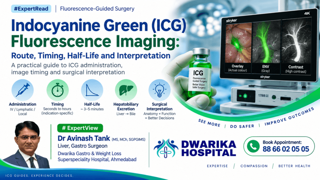

A practical guide to ICG administration, image timing and surgical interpretation
#ExpertRead | Fluorescence-Guided Surgery
Indocyanine green (ICG) has evolved from a vascular diagnostic dye into one of the most useful fluorescence-guided tools in modern surgery. When combined with near-infrared (NIR) imaging systems such as Stryker SPY, ICG can provide real-time information about blood flow, tissue perfusion, biliary anatomy, lymphatic drainage and selected anatomical structures.
The most important concept, however, is simple:
ICG timing is indication-dependent. The same injection does not produce the same useful image at every point in time.
For vascular imaging, the useful signal appears within seconds. For biliary imaging, the surgeon deliberately waits for hepatic uptake and biliary excretion. For lymphatic mapping, the route of administration determines how quickly fluorescence reaches the lymphatic channels and nodes.
Understanding the pharmacokinetics of ICG therefore becomes essential for correct image interpretation.
1. What is ICG?
Indocyanine green (ICG) is a water-soluble near-infrared fluorescent dye that binds strongly to plasma proteins after intravenous administration.
It absorbs and emits light in the near-infrared spectrum, allowing fluorescence to be detected by dedicated NIR imaging systems.
Its major pharmacological characteristics are:
- Rapid intravascular distribution
- Very high plasma protein binding
- Predominantly hepatic uptake
- Excretion into bile
- Minimal metabolism
- Very short plasma half-life
- Repeat dosing can therefore be possible during surgery when clinically appropriate
Current prescribing information supports ICG fluorescence imaging for vessels/blood flow/tissue perfusion, extrahepatic biliary ducts and selected lymphatic mapping applications, with dose and route depending on the indication. (DailyMed)
2. Why ICG is valuable in surgery
ICG provides information that conventional white-light imaging cannot directly provide.
Vascular surgery / perfusion
ICG can demonstrate:
- Arterial inflow
- Tissue perfusion
- Anastomotic perfusion
- Bowel viability
- Gastric conduit perfusion
- Flap perfusion
- Microvascular reconstruction
Hepatobiliary surgery
ICG fluorescence can help identify:
- Common bile duct
- Common hepatic duct
- Cystic duct
- Biliary confluence
- Extrahepatic biliary anatomy
This is particularly useful during difficult laparoscopic or robotic cholecystectomy.
Lymphatic surgery
Depending on the indication and route, ICG can identify:
- Lymphatic channels
- Sentinel lymphatic drainage
- Regional lymph nodes
The major advantage
ICG is not simply an anatomical imaging dye.
It can provide functional information:
“Is blood reaching this tissue?”
rather than merely:
“What does this tissue look like?”
That distinction is particularly important when assessing anastomoses and tissue viability.
3. The most important concept: ICG has a very short plasma half-life
The plasma half-life of ICG is approximately 3–5 minutes, although published pharmacokinetic estimates vary somewhat according to methodology and clinical circumstances.
This short half-life has an important surgical implication:
IV ICG is primarily a dynamic test rather than a static stain.
After intravenous injection, the concentration in circulating blood falls rapidly.
Therefore, the fluorescence seen immediately after injection is fundamentally different from the fluorescence seen later.
Early after injection
Blood-pool fluorescence dominates.
This is ideal for:
- Arterial inflow
- Vessel identification
- Perfusion assessment
- Anastomotic evaluation
Later after injection
ICG is progressively removed from the circulation by the liver and excreted into bile.
Consequently:
Blood fluorescence ↓
while:
Biliary fluorescence ↑
This is why a short plasma half-life does not mean that ICG fluorescence disappears from the operation.
The dye has moved from the vascular compartment toward the hepatobiliary system.
4. ICG pharmacokinetics: why the surgeon should care
The simplified pathway is:
Injection
↓
Bloodstream
↓
Protein binding
↓
Hepatic uptake
↓
Biliary excretion
↓
Bile duct fluorescence
↓
Intestinal/biliary elimination
ICG is predominantly eliminated through the hepatobiliary route rather than renal excretion.
Therefore, the timing of imaging must be interpreted according to where the dye is expected to be at that moment.
5. Route of administration determines what you see
There are three major practical routes.
| Route | Main purpose | Expected imaging |
|---|---|---|
| IV | Vascular/perfusion | Seconds |
| IV | Biliary mapping | ≥45 min; often longer in clinical practice |
| Interstitial / intradermal / subcutaneous | Lymphatic mapping | Minutes; depends on injection site and lymphatic drainage |
| Direct/local biliary administration | Selected specialised applications | Immediate/local fluorescence |
The route should therefore never be separated from the indication.
6. IV ICG for vascular and perfusion imaging
For vascular imaging, ICG is administered as an intravenous bolus, followed by an appropriate saline flush according to the product/device protocol.
Current prescribing information states that fluorescence should become visible in blood vessels approximately 5–15 seconds after injection. (DailyMed)
Practical sequence
T = 0
→ IV ICG injection
T = 5–15 sec
→ vascular fluorescence appears
T = 10–60 sec
→ evaluate arterial inflow and tissue perfusion
Following correction
→ repeat assessment can be performed when clinically appropriate
What should the surgeon look for?
Do not simply ask:
“Is it fluorescent?”
Ask:
“How rapidly, how uniformly and how strongly does fluorescence reach the tissue?”
This changes ICG from a picture-taking exercise into a functional perfusion assessment.
7. Anastomotic assessment
ICG can be particularly valuable immediately before committing to an anastomosis.
Continue Reading — Free
Get the complete expert guide from Dr. Avinash Tank.
Free, always. No payment, no spam.
Continue Reading — Free
Get the complete expert guide from Dr. Avinash Tank.
Free, always. No payment, no spam — just the complete guide sent straight to your screen.
For example:
Proposed bowel transection line
↓
IV ICG
↓
5–15 seconds
↓
Observe perfusion
↓
Change transection line if necessary
↓
Repeat ICG assessment
↓
Construct anastomosis
↓
Optional postoperative/anastomotic reassessment
The key advantage is that ICG can provide real-time functional information before an irreversible surgical decision.
However, fluorescence should complement—not replace—clinical judgement, anatomy, haemodynamic assessment and established surgical principles.
8. ICG for biliary fluorescence cholangiography
This is where timing becomes particularly important.
After IV administration, ICG reaches the liver, is taken up by hepatocytes and is excreted into bile.
The bile ducts therefore become fluorescent later than the blood vessels.
Current prescribing information recommends:
2.5 mg IV ≥45 minutes before surgery
for extrahepatic biliary duct visualization, with fluorescence expected in the biliary tree within approximately 45 minutes. (DailyMed)
Why not inject immediately before looking for the bile duct?
Because immediately after IV injection there is substantial fluorescence within:
- Blood
- Liver
- Vascular structures
This can produce a high background signal.
The objective of delayed biliary imaging is to allow:
Blood-pool fluorescence ↓
while:
Biliary fluorescence ↑
thereby improving the bile duct-to-liver contrast.
9. Timing of ICG for cholecystectomy
There is no universally accepted single timing protocol for fluorescence cholangiography.
Published studies have used administration intervals ranging from approximately 15 minutes to 24 hours before surgery, with substantial variation in dose and timing. A 2026 systematic analysis of 54 studies involving 3,070 patients found IV administration to be the predominant route, while emphasizing the continuing heterogeneity of protocols. (PubMed)
Expert consensus has favoured giving ICG at least 45 minutes before the procedure in order to reduce intense hepatic background fluorescence. (PubMed)
Some clinical studies have investigated administration on the day before surgery to further improve bile duct-to-liver contrast. (PubMed)
Practical interpretation
For routine biliary fluorescence:
Short interval
→ more blood/liver background
≥45 min
→ useful biliary fluorescence with reduced blood-pool component
Several hours / overnight
→ potentially improved duct-to-liver contrast in some protocols
But:
Longer is not automatically better.
The optimal timing depends on dose, liver function, inflammation, body habitus, equipment, indication and the fluorescence characteristics of the imaging system.
10. Difficult cholecystectomy: what ICG can and cannot do
ICG fluorescence can help identify biliary structures before and during dissection.
Potential advantages include:
- Real-time visualization
- No ionising radiation
- No need for duct cannulation
- Repeated imaging
- Improved anatomical orientation
- Assistance in obese patients
- Assistance in inflammatory cases
A systematic review found that ICG fluorescence cholangiography improves visualization of biliary anatomy, although there remains heterogeneity regarding dose, timing and administration protocols. (PubMed)
Importantly:
ICG fluorescence is an adjunct—not a substitute for sound surgical anatomy and critical-view principles.
Severe inflammation, impacted stones, previous biliary intervention and other factors can reduce visualization quality. (PubMed)
11. Lymphatic mapping: a different route, a different clock
Lymphatic imaging is fundamentally different from IV perfusion imaging.
ICG is injected intradermally, subcutaneously or interstitially, depending on the procedure.
The dye then travels through lymphatic channels toward regional lymph nodes.
For example, the current prescribing information describes:
Cervical/uterine lymphatic mapping
→ 5 mg interstitially
→ four injections
→ superficial and deep cervical injection sites
→ lymphatic vessels and nodes should begin to become visible within approximately 1 minute. (DailyMed)
The exact technique, concentration and dose are indication-specific.
Therefore:
Never transfer an IV vascular dose directly to a lymphatic protocol.
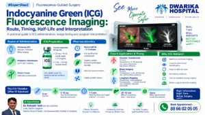
12. A practical “ICG clock” for surgeons
0 seconds — IV injection
ICG enters bloodstream.
5–15 seconds
Vascular fluorescence
Best for:
- Arterial inflow
- Vessel visualization
- Tissue perfusion
- Anastomotic assessment
(DailyMed)
1–5 minutes
The circulating signal begins to decline because of the short plasma half-life.
Useful interpretation:
Dynamic perfusion rather than static staining.
30–45+ minutes
ICG has undergone hepatic uptake and biliary excretion.
Biliary fluorescence becomes clinically useful.
The current labelled protocol specifies ≥45 minutes for extrahepatic biliary imaging. (DailyMed)
Several hours
Biliary fluorescence can remain useful, depending on the clinical situation and imaging system.
However, signal quality is influenced by:
- Hepatic function
- Dose
- Timing
- Inflammation
- Tissue thickness
- Obesity
- Liver background
- Imaging distance
- Camera angle
- Device sensitivity
Hours later
The clinical interpretation should focus on where the ICG has moved, rather than assuming that the original intravascular signal remains unchanged.
13. Why “half-life” should not be confused with “imaging duration”
This is one of the most important teaching points.
Plasma half-life ≠ fluorescence duration
A drug with a 3–5 minute plasma half-life does not necessarily produce only 3–5 minutes of useful surgical fluorescence.
Why?
Because ICG is moving between physiological compartments.
Blood
→
Liver
→
Bile
→
Intestine
The surgeon is observing different compartments at different times.
Therefore:
The timing of the image determines the physiological question being answered.
14. Route × timing × question
A useful mental model is:
| Clinical question | Route | Approximate imaging window |
|---|---|---|
| Is blood reaching the tissue? | IV | 5–15 sec onward |
| Is the anastomosis perfused? | IV | Seconds to minutes |
| Where is the bile duct? | IV | ≥45 min; often longer depending on protocol |
| Where are the lymphatics? | Intradermal/interstitial | ~1 min onward; procedure dependent |
| Is tissue perfusion improved after correction? | IV repeat dose | Seconds after reinjection |
The exact protocol should always follow the specific ICG product label, imaging system and surgical indication.
15. How to interpret a “weak” ICG image
A weak image does not automatically mean poor perfusion.
Possible explanations include:
Patient factors
- Obesity
- Thick tissue
- Poor cardiac output
- Low arterial inflow
- Hypotension
- Liver dysfunction for biliary applications
Surgical factors
- Severe inflammation
- Oedema
- Haematoma
- Tissue depth
- Excessive distance from camera
- Incorrect camera angle
Technical factors
- Insufficient dose
- Incorrect timing
- Excessive ambient/background signal
- Device settings
- Poor exposure geometry
A systematic review of fluorescence cholangiography demonstrated that fluorescence intensity is affected by several variables, including obesity and inflammation. (PubMed)
Therefore:
Interpret the fluorescence image in context—not as a binary “green = good / no green = bad” test.
16. ICG with modern NIR systems
Modern fluorescence platforms can present ICG information in different visualization modes.
For example, systems may provide:
- White-light anatomy with fluorescence overlay
- Grayscale fluorescence visualization
- High-contrast fluorescence display
The purpose is not merely aesthetic.
Different visualization modes can change the surgeon’s ability to distinguish a weak fluorescence signal from background anatomy or liver fluorescence.
The most important principle remains:
The imaging system detects the fluorescence; the surgeon must understand the physiology behind the fluorescence.
17. Practical checklist before injecting ICG
Before administration, ask five questions:
1. What am I trying to see?
Perfusion? Bile duct? Lymphatics?
2. What is the correct route?
IV or local/interstitial?
3. When should I inject?
Seconds before vascular assessment?
≥45 minutes before biliary imaging?
Procedure-specific timing for lymphatic mapping?
4. What should I expect to see?
Know the expected fluorescence window before injecting.
5. What does a weak signal mean?
Consider:
Timing + dose + physiology + tissue depth + inflammation + equipment
before concluding that the anatomy or perfusion is abnormal.
18. The surgeon’s takeaway
ICG fluorescence is best understood as a time-dependent physiological imaging technique.
Its short plasma half-life is not a limitation—it is one of its advantages.
For vascular imaging:
Inject → seconds → assess perfusion
For biliary imaging:
Inject → hepatic uptake → biliary excretion → wait → identify ducts
For lymphatic imaging:
Local injection → lymphatic transport → visualize channels/nodes
The same molecule therefore answers very different surgical questions depending on route, dose, timing and imaging modality.
The simplest rule to remember
ICG is not simply a dye. It is a dynamic tracer.
Seconds = blood flow.
Tens of minutes = biliary mapping.
Local injection + minutes = lymphatic mapping.
And the most important interpretive principle:
Always interpret the fluorescence according to where the ICG should physiologically be at that moment.
Key references
- SPY AGENT GREEN / Indocyanine Green prescribing information. Current labeling specifies 5–15-second vascular fluorescence and ≥45-minute biliary imaging after IV administration. (DailyMed)
- Systematic review of ICG near-infrared fluorescence in laparoscopic cholecystectomy. Demonstrates benefits for biliary visualization while highlighting heterogeneity in dose and timing. (PubMed)
- International Delphi consensus on ICG fluorescence during laparoscopic cholecystectomy. Supports NIR fluorescence for biliary visualization and discusses dose/timing considerations. (PubMed)
- Systematic review and ex-vivo evaluation of ICG fluorescence cholangiography. Examines factors affecting fluorescence intensity and image quality. (PubMed)
- 2026 systematic analysis of ICG fluorescence during cholecystectomy. Reviews route, dose, timing and detection outcomes across 54 studies and 3,070 patients. (PubMed)
Bottom line
ICG timing should be planned backward from the surgical question.
Perfusion → inject immediately before assessment.
Biliary anatomy → inject sufficiently early, with ≥45 minutes being the labelled approach for extrahepatic biliary imaging.
Lymphatic mapping → use the procedure-specific local/interstitial protocol.
Never interpret ICG fluorescence without considering the time elapsed since injection.
This can also be converted into a 6–8 slide “ICG: The Surgeon’s Clock” presentation, with a single timeline graphic from 0 seconds → 15 seconds → 45 minutes → several hours, which would work particularly well for a surgical video presentation.
Related patient videos
 DWARIKA HOSPITALVerified Patient Story
Verified
DWARIKA HOSPITALVerified Patient Story
Verified  DWARIKA HOSPITALVerified Patient Story
Verified
DWARIKA HOSPITALVerified Patient Story
Verified  DWARIKA HOSPITALVerified Patient Story
Verified
DWARIKA HOSPITALVerified Patient Story
Verified 
Related treatments & services
A complete, personalised weight-loss programme — surgery where needed, plus nutrition, medical and lifestyle support for lasting results.
View service → 360 Diabetes ReversalA complete programme to reverse Type-2 diabetes — combining metabolic surgery where suitable with diet, medical and lifestyle care.
View service → Gallbladder Stone
Minimally invasive removal of the gallbladder (laparoscopic cholecystectomy) — the definitive, day-care treatment for painful gallstones.
View service →Was this guide helpful?
Super-specialist GI, bariatric & cancer surgeon. SGPGIMS (India's premier GI centre) + advanced training in Japan & South Korea. Read full profile →
Last reviewed: July 2026