

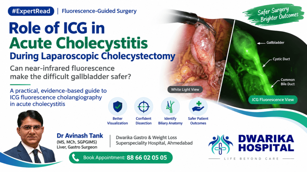

Can near-infrared fluorescence make the difficult gallbladder safer?
A practical, evidence-based guide to ICG fluorescence cholangiography in acute cholecystitis
The clinical problem
Acute cholecystitis is not simply a routine laparoscopic cholecystectomy performed urgently.
Inflammation produces edema, tissue friability, adhesions, distorted Calot’s triangle anatomy and fibrosis, making identification of the cystic duct, common hepatic duct and common bile duct progressively more difficult.
The fundamental safety principle remains the Critical View of Safety (CVS). ICG fluorescence should therefore be regarded as an adjunct to anatomical dissection—not a replacement for CVS.
This distinction is particularly important because the evidence demonstrates that ICG improves visualization, but has not yet conclusively demonstrated a reduction in bile duct injury in randomized trials. (PubMed)
What exactly does ICG add?
Indocyanine green (ICG) is administered intravenously and rapidly binds to plasma proteins. It is taken up by hepatocytes and excreted into bile.
When illuminated with near-infrared light, ICG produces fluorescence that allows the surgeon to visualize bile-containing structures.
During laparoscopic cholecystectomy, this can potentially identify:
- Cystic duct
- Common hepatic duct
- Common bile duct
- Cystic duct–CBD junction
- Confluence of the hepatic ducts in favourable circumstances
The key advantage is that the information is available in real time without cannulating the cystic duct and without X-ray exposure.
A clinical study reported visualization of the cystic duct, CBD and CHD before dissection in 91%, 79% and 53% of patients respectively. (PubMed)
Why could ICG be particularly useful in acute cholecystitis?
1. Inflammation changes anatomy
The surgeon may encounter:
“Where is the cystic duct?”
ICG can provide an additional anatomical map before aggressive dissection begins.
This is potentially valuable when the inflammatory mass makes conventional identification difficult.
2. It can provide information before dissection
Unlike conventional cholangiography, fluorescence can be observed before entering Calot’s triangle.
This may help the surgeon understand the relationship between the cystic duct and the extrahepatic biliary tree before committing to clipping or division.
3. It may increase surgical confidence
The 2025 WSES international consensus specifically addressed emergency surgery and concluded that ICG cholangiography enhances biliary-tree visualization and can assist dissection toward the Critical View of Safety in acute cholecystitis. (PubMed Central (PMC))
What does the evidence actually show?
This is where the distinction between better visualization and better outcomes becomes important.
Evidence supporting visualization
A 2025 systematic review and meta-analysis restricted to randomized controlled trials included 8 studies and 1,586 patients.
ICG fluorescence significantly improved the odds of identifying the common bile duct:
OR 4.08 (95% CI 1.77–9.41)
However, statistically significant improvements were not demonstrated for bile duct injury, cystic duct identification or common hepatic duct identification. (PubMed)
An earlier meta-analysis of 22 studies involving 3,457 patients similarly found improved identification of the cystic duct and CBD, shorter identification time and lower conversion rates, although substantial heterogeneity was present. (PubMed)
But does ICG prevent bile duct injury?
We cannot yet say that with certainty.
A randomized trial specifically examining emergency laparoscopic cholecystectomy included 92 patients with acute cholecystitis.
ICG fluorescence did not significantly reduce conversion, operative time, complications or bile leak compared with conventional surgery.
The authors concluded that routine use was questionable, while suggesting that ICG may be particularly useful in difficult cholecystectomy. (PubMed)
Similarly, the 2025 RCT-only meta-analysis did not demonstrate a statistically significant reduction in bile duct injury. (PubMed)
This is partly a statistical problem: bile duct injury is fortunately uncommon, meaning enormous sample sizes are required to demonstrate a meaningful reduction.
The important 2026 perspective
More recent evidence is encouraging.
A 2026 systematic review and meta-analysis of difficult laparoscopic cholecystectomy included 1,069 patients and reported that ICG fluorescence was associated with shorter operative time and hospital stay, with a lower observed conversion-to-open rate. However, only one randomized trial was included, meaning the findings should be interpreted cautiously. (PubMed)
An even larger 2026 national cohort involving 1.27 million cholecystectomy patients found that ICG use was associated with lower CBD injury, subsequent biliary intervention and conversion rates after adjustment. Importantly, this was an observational study—not randomized evidence—so association cannot establish causation. (PubMed)
Continue Reading — Free
Get the complete expert guide from Dr. Avinash Tank.
Free, always. No payment, no spam.
Continue Reading — Free
Get the complete expert guide from Dr. Avinash Tank.
Free, always. No payment, no spam — just the complete guide sent straight to your screen.
The message for surgeons:
ICG clearly improves visualization.
Whether it independently prevents bile duct injury remains less certain.
That is an important distinction when counselling surgeons about the technology.
ICG in the difficult acute gallbladder
This may actually be the most logical application.
Consider:
- Severe acute cholecystitis
- Dense adhesions
- Contracted gallbladder
- Impacted neck stone
- Mirizzi-type anatomy
- Short or parallel cystic duct
- Distorted Calot’s triangle
- Gangrenous gallbladder
- Previous episodes of cholecystitis
- Unexpected anatomy
- Difficult identification of the cystic duct
In these situations, the question is not:
“Can ICG replace dissection?”
The better question is:
“Can ICG give me additional anatomical information before I make the next irreversible move?”
That is where fluorescence potentially adds the greatest value.
Practical intraoperative workflow
Step 1 — Administer ICG
Intravenous administration is the most commonly used approach.
However, dose and timing are not completely standardized.
A randomized dose-finding study evaluated administration approximately 30 minutes before surgery and found that 0.1 mg provided favourable bile duct-to-liver fluorescence ratios compared with higher doses in its specific imaging system and protocol. (PubMed)
Therefore, surgeons should follow the dose, concentration, timing and imaging-system recommendations of the specific ICG product and fluorescence platform rather than universally adopting one published dose.
Step 2 — Start with white light
ICG should not replace conventional anatomical orientation.
Begin with:
White light → exposure → decompression → traction → anatomical landmarks
Then switch to fluorescence.
Step 3 — Map the biliary anatomy
Before significant dissection, identify:
CBD → CHD → cystic duct → gallbladder
The fluorescence image should be interpreted together with the white-light anatomy.
Step 4 — Dissect toward CVS
The surgeon should continue to obtain the Critical View of Safety.
ICG can help answer:
- Is this structure the cystic duct?
- Where is the CBD?
- Is the CBD running close to the operative field?
- Is the suspected cystic duct actually entering the gallbladder?
- Has inflammation distorted the expected anatomy?
The “ICG pause” before clipping
A useful practical concept is to introduce an ICG safety pause immediately before clipping or dividing the cystic duct.
Ask three questions:
1. What is this structure?
2. Where is the CBD/CHD?
3. Do the fluorescence anatomy and surgical anatomy agree?
If they do not agree:
STOP.
Do not proceed simply because the structure “looks like” the cystic duct.
This is particularly relevant in severe inflammation, where the consequences of a mistaken assumption can be catastrophic.
What ICG cannot do
ICG has important limitations.
It does NOT:
- Demonstrate the Critical View of Safety by itself
- Replace careful dissection
- Reliably identify structures buried under thick inflammatory tissue
- Replace intraoperative cholangiography when ductal stones or detailed ductal anatomy need evaluation
- Reliably identify vascular anatomy
- Guarantee prevention of bile duct injury
- Replace bailout procedures when CVS cannot be achieved
The fluorescence signal can also be affected by liver background fluorescence, excessive ICG dose, timing of administration, obesity, inflammation and tissue thickness.
ICG versus conventional IOC
This is an important practical distinction.
ICG fluorescence
Advantages
- No X-ray
- No cystic duct cannulation
- Real-time imaging
- Rapid
- Repeatable
- Can be used before dissection
- Provides continuous visual guidance
Conventional intraoperative cholangiography
Advantages
- Provides a ductal contrast study
- Better assessment of ductal continuity
- Can demonstrate filling defects
- Can provide more comprehensive biliary anatomy
The 2025 SAGES guideline conditionally favours IOC over ICG fluorescence imaging for benign biliary disease, reflecting the current certainty of evidence rather than suggesting that ICG is clinically useless. (PubMed)
Therefore:
ICG and IOC should be viewed as complementary technologies rather than interchangeable techniques.
What does WSES recommend?
The position of the emergency-surgery community is particularly relevant to acute cholecystitis.
The WSES international consensus position paper states that ICG cholangiography can enhance visualization of the biliary tree, guide dissection toward CVS and potentially improve confidence during emergency laparoscopic cholecystectomy.
Importantly, the panel recommends ICG cholangiography during laparoscopic cholecystectomy for acute cholecystitis when equipment and expertise are available, while acknowledging that the underlying evidence is moderate/low level. (PubMed Central (PMC))
This is therefore a strong recommendation with an evidence base that is not equivalent to high-quality RCT evidence—an important nuance.
A practical algorithm
Acute cholecystitis → Laparoscopic cholecystectomy
↓
Administer ICG according to local protocol
↓
White-light anatomical assessment
↓
Near-infrared fluorescence mapping
↓
Identify CBD/CHD + cystic duct
↓
Progressive dissection
↓
Critical View of Safety
↓
ICG + white-light confirmation
↓
Clip and divide cystic duct
↓
If CVS cannot be achieved → STOP
↓
Consider bailout strategy
- Subtotal cholecystectomy
- Fundus-first approach in selected circumstances
- Conversion when appropriate
- Intraoperative cholangiography
- Additional imaging/consultation according to the clinical situation
The most important surgical principle
ICG should be an adjunct, not a permission slip.
A fluorescent cystic duct does not automatically mean that the anatomy is safe.
The surgeon must still establish the relationship between:
Gallbladder → cystic duct → CHD → CBD
and obtain the Critical View of Safety before division.
If the anatomy remains unclear despite fluorescence:
Do not dissect harder. Change the strategy.
That may mean subtotal cholecystectomy, conversion, IOC or another bailout technique.
Expert takeaway
The role of ICG in acute cholecystitis is best understood as real-time anatomical augmentation.
It can:
✔ Improve visualization of extrahepatic bile ducts
✔ Provide anatomical information before and during dissection
✔ Potentially shorten identification of biliary structures
✔ Be particularly useful in difficult/inflamed Calot’s triangle
✔ Avoid radiation and cystic duct cannulation
But:
✘ It does not replace Critical View of Safety
✘ It does not guarantee prevention of bile duct injury
✘ It cannot reliably resolve every difficult anatomy
✘ Current randomized evidence has not conclusively demonstrated reduction in bile duct injury
Bottom line
In acute cholecystitis, ICG is best viewed as an additional pair of eyes—not as a substitute for surgical judgment.
The strongest current evidence supports its ability to improve biliary visualization, while evidence for reducing clinically important outcomes such as bile duct injury remains evolving.
For the surgeon facing a severely inflamed Calot’s triangle, however, the practical value may be considerable:
See the anatomy → understand the anatomy → achieve CVS → confirm before dividing.
And when anatomy remains uncertain:
ICG should make the surgeon more cautious—not more confident.
That may ultimately be its greatest contribution to safer emergency laparoscopic cholecystectomy.
Key PubMed references
- Calabrese EC, et al. SAGES 2025 guidelines for fluorescence image-guided surgery using indocyanine green. Surg Endosc. 2025;39:7935-7957. PMID: 41249539. (PubMed)
- Kumar SK, et al. SAGES guidelines for the use of intraoperative imaging of the common bile duct. Surg Endosc. 2025. PMID: 41044285. (PubMed)
- Pimentel T, et al. Indocyanine green fluorescent cholangiography in laparoscopic cholecystectomy: systematic review and meta-analysis with trial sequential analysis of randomized controlled trials. Surgery. 2025;181:109149. PMID: 39891966. (PubMed)
- Seretis C, et al. Routine use of ICG to enhance operative safety in emergency laparoscopic cholecystectomy: a randomized controlled trial. PMID: 35194663. (PubMed)
- Huang Y, et al. Real-time fluorescent cholangiography with indocyanine green in laparoscopic cholecystectomy: randomized controlled trial to establish optimal ICG dose. Surg Today. 2023. PMID: 35920936. (PubMed)
- Dip F, et al. Does near-infrared fluorescent cholangiography with indocyanine green reduce bile duct injuries and conversions to open surgery? Surgery. 2021. PMID: 33478756. (PubMed)
- Pesce A, et al. Utility of fluorescent cholangiography during laparoscopic cholecystectomy: a systematic review. World J Gastroenterol. 2015;21:7877-7883. PMID: 26167088. (PubMed)
- WSES. Indocyanine green fluorescence-guided surgery in the emergency setting: international consensus position paper. (PubMed Central (PMC))
- WSES. 2020 guidelines for detection and management of bile duct injury during cholecystectomy. (PubMed Central (PMC))
- Tang K, et al. Evaluating the safety and efficacy of ICG fluorescence imaging for difficult laparoscopic cholecystectomy: systematic review and meta-analysis. Am Surg. 2026;92:1374-1383. PMID: 41289059. (PubMed)
Related patient videos
 DWARIKA HOSPITALVerified Patient Story
Verified
DWARIKA HOSPITALVerified Patient Story
Verified  DWARIKA HOSPITALVerified Patient Story
Verified
DWARIKA HOSPITALVerified Patient Story
Verified  DWARIKA HOSPITALVerified Patient Story
Verified
DWARIKA HOSPITALVerified Patient Story
Verified 
Related treatments & services
A complete, personalised weight-loss programme — surgery where needed, plus nutrition, medical and lifestyle support for lasting results.
View service → 360 Diabetes ReversalA complete programme to reverse Type-2 diabetes — combining metabolic surgery where suitable with diet, medical and lifestyle care.
View service → Gallbladder Stone
Minimally invasive removal of the gallbladder (laparoscopic cholecystectomy) — the definitive, day-care treatment for painful gallstones.
View service →Was this guide helpful?
Super-specialist GI, bariatric & cancer surgeon. SGPGIMS (India's premier GI centre) + advanced training in Japan & South Korea. Read full profile →
Last reviewed: July 2026