Reading Time: 13 minutes
Large Hiatus Hernia with Severe GERD Successfully Treated by Advanced Laparoscopic Anti-Reflux Surgery | Dr Avinash Tank
A Clinical Case Library by Dr. Avinash Tank
Clinical Case Overview
Some operations begin exactly as expected.
Others reveal an unexpected challenge that tests the surgeon’s judgement, adaptability, and technical precision.
This case belonged to the second category.
A woman in her late sixties sought medical attention because every meal had become uncomfortable.
Food repeatedly came back into her mouth, persistent acidity disturbed her daily routine, and bloating after meals made eating an unpleasant experience.
Although medicines provided temporary relief, her symptoms continued to worsen over the preceding weeks, suggesting that the problem was more than excess stomach acid alone.
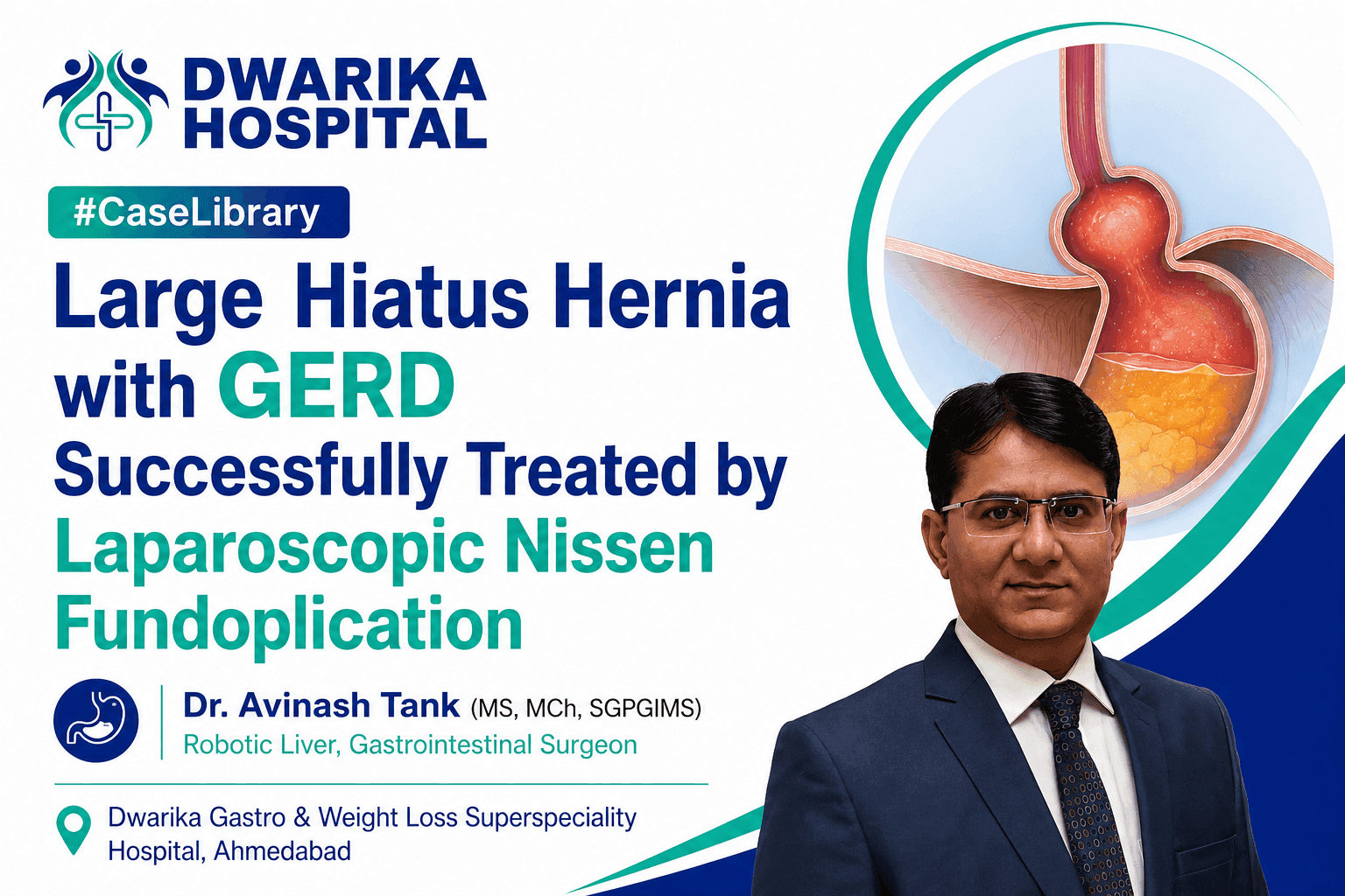
Further evaluation confirmed gastroesophageal reflux disease (GERD) associated with a hiatus hernia.
However, the most interesting finding was not discovered during endoscopy—it was identified only after the laparoscopic camera entered the abdomen.
A large fatty mass surrounding the gastroesophageal junction added an unexpected layer of complexity to what initially appeared to be a routine anti-reflux operation.
This case demonstrates how careful surgical planning, meticulous laparoscopic technique, and sound intraoperative judgement can safely address multiple problems during a single minimally invasive procedure.
Quick Facts
Patient: Woman in her late sixties
Primary Symptoms:
- Persistent food regurgitation
- Acid reflux
- Upper abdominal pain
- Bloating after meals
- Excessive acidity
Final Diagnosis:
- Gastroesophageal Reflux Disease (GERD)
- Sliding Hiatus Hernia
- Fatty mass at the Gastroesophageal Junction (suspected lipoma)
Procedure Performed:
- Laparoscopic reduction of hiatus hernia
- Complete excision of fatty tissue at the gastroesophageal junction
- Posterior cruroplasty
- 360° floppy Nissen fundoplication
Outcome:
Successful minimally invasive surgery with an uneventful recovery and stable discharge.
Why This Case Matters
Many people believe persistent acidity is simply the result of “too much stomach acid.”
In reality, chronic reflux is often caused by a mechanical problem rather than a chemical one.
A healthy person has a natural anti-reflux valve where the oesophagus joins the stomach. This valve works together with the diaphragm to prevent stomach contents from flowing backwards.
When a hiatus hernia develops, part of the stomach slides upward through the diaphragm into the chest. As the normal anatomy becomes distorted, the anti-reflux mechanism weakens. Acid, food, and digestive juices can then travel back into the oesophagus, producing heartburn, regurgitation, chest discomfort, and bloating.
Medicines can reduce acid production, but they cannot reposition the stomach or repair the enlarged opening in the diaphragm. Understanding this difference is crucial when deciding whether surgery may provide a more durable solution.
The Patient’s Journey
For almost two months, eating had become increasingly difficult.
Meals were followed by burning discomfort behind the chest, repeated regurgitation of food, abdominal pain, and troublesome bloating. Over the previous fifteen days these symptoms became noticeably worse, despite ongoing medical treatment. Rather than improving, everyday activities such as eating, sleeping, and socialising were increasingly affected.
This pattern is important. Progressive symptoms despite appropriate medication often suggest that the underlying problem is structural rather than simply excessive acid production.
Recognising this possibility, the patient sought specialist evaluation.
Understanding the Symptoms
Each symptom reflected a different aspect of the underlying disease.
Food Regurgitation
Food returning into the mouth usually indicates failure of the natural valve between the stomach and oesophagus rather than overproduction of acid alone.
Persistent Heartburn
Acid repeatedly irritating the lining of the oesophagus produces the familiar burning sensation known as heartburn.
Bloating After Meals
When the stomach is displaced into the chest, normal movement of food and gas can become disturbed, leading to fullness and bloating.
Upper Abdominal Pain
Stretching of the hiatus, chronic inflammation, and repeated reflux episodes may all contribute to discomfort in the upper abdomen.
Taken together, these symptoms strongly suggested that correcting the anatomy—not merely suppressing acid—would ultimately be required.
Medical Background
Like many patients requiring upper gastrointestinal surgery, this patient also had long-standing medical conditions including hypertension, type 2 diabetes mellitus, and dyslipidaemia. These conditions had been treated medically for many years.
Although these illnesses did not directly cause the hiatus hernia, they influenced perioperative planning. Careful assessment of cardiovascular fitness, blood glucose control, and overall medical stability was therefore completed before surgery was considered.
Clinical Assessment
Physical examination revealed tenderness over the upper abdomen, supporting the history of ongoing upper gastrointestinal pathology. Importantly, there was no history of alcohol use, tobacco consumption, or significant allergies that would complicate perioperative management.
At this stage, several possibilities could explain the patient’s symptoms, including severe GERD, gastritis, peptic ulcer disease, or a significant hiatus hernia. The next step was to identify the exact anatomical problem.
Diagnostic Work-up
Upper gastrointestinal endoscopy provided the key information.
It demonstrated:
- Gastroesophageal reflux disease (GERD)
- A 3 cm sliding hiatus hernia
- Diffuse gastritis
Routine blood investigations showed satisfactory haemoglobin, kidney function, liver function, and coagulation profile. Chest X-ray was within normal limits, while echocardiography confirmed preserved heart function suitable for general anaesthesia.
These investigations answered two critical questions:
- What was causing the symptoms?
- Was the patient fit to undergo laparoscopic surgery?
Both questions could now be answered confidently.
Clinical Reasoning
The central question was not whether the patient had reflux.
The real question was why the reflux persisted despite treatment.
The endoscopy demonstrated that the lower oesophageal sphincter was no longer functioning within its normal anatomical position because part of the stomach had migrated through the diaphragmatic opening.
This explained why medications had failed to provide lasting relief.
Suppressing acid could reduce irritation, but it could not restore the anatomy responsible for preventing reflux.
Therefore, treatment needed to address the structural defect itself rather than simply its symptoms.
Why Surgery Was Recommended
Not every patient with GERD requires an operation.
Many improve with lifestyle modification and medication.
However, surgery becomes an important option when:
- Symptoms remain troublesome despite optimal medical therapy.
- A significant hiatus hernia is present.
- Food regurgitation affects quality of life.
- The underlying anatomy has become abnormal.
In this case, the patient’s persistent symptoms, documented hiatus hernia, and failure of conservative treatment made laparoscopic anti-reflux surgery the most logical treatment strategy.
The aim was not simply to stop acid production but to restore the normal relationship between the stomach, oesophagus, and diaphragm, thereby rebuilding the body’s natural anti-reflux mechanism.
At this stage, the operation appeared straightforward. However, the surgical team would soon discover an unexpected finding that transformed this into a much more technically demanding procedure.
Inside the Operation: Surgical Planning, Operative Findings and Technical Challenges
Surgical Planning
Every successful operation begins long before the first incision.
For this patient, the surgical objective was clear: restore the normal anatomy of the stomach and oesophagus, repair the widened diaphragmatic opening, and recreate the natural anti-reflux barrier. However, achieving these goals safely required careful preoperative planning.
Because the patient had long-standing hypertension, diabetes mellitus and dyslipidaemia, medical fitness was obtained before surgery. Anaesthetic assessment confirmed that she could safely undergo general anaesthesia, while critical care support was arranged for postoperative monitoring considering her age and associated medical conditions.
The operation was planned as a laparoscopic procedure, offering the advantages of smaller incisions, reduced postoperative pain, earlier mobilisation, faster recovery and lower wound-related complications compared with conventional open surgery.
Although the diagnosis appeared straightforward, experienced surgeons know that the true complexity of a hiatus hernia often becomes apparent only after entering the abdomen.
Why Laparoscopic Surgery Was the Preferred Choice
A hiatus hernia is fundamentally an anatomical problem.
The stomach has moved into a position where it should not be, weakening the body’s natural anti-reflux mechanism. Simply reducing stomach acid does not correct this displacement.
Laparoscopic surgery allows the surgeon to:
- Magnify delicate anatomical structures.
- Safely separate the oesophagus from surrounding tissues.
- Return the stomach to its normal position.
- Repair the enlarged opening in the diaphragm.
- Construct a new anti-reflux valve using the patient’s own stomach.
Because the operation is performed through small incisions using a high-definition camera, patients generally experience faster recovery while still receiving a durable anatomical repair.
Entering the Abdomen
After induction of general anaesthesia, the patient was placed in the French position, which provides excellent access to the upper abdomen during advanced laparoscopic surgery.
A pneumoperitoneum was created using a Veress needle, followed by placement of five laparoscopic ports under direct vision. The laparoscope immediately provided a magnified view of the upper abdomen, allowing systematic assessment before beginning definitive surgery.
This first inspection confirmed that the operation would be more complex than expected.
What We Discovered During Surgery
The laparoscopic camera revealed a wide oesophageal hiatus, much larger than normal.
Instead of remaining securely within the abdominal cavity, the upper portion of the stomach had migrated through this enlarged opening into the lower chest. This abnormal position explained why the patient had experienced persistent reflux despite medication. The natural anti-reflux valve could no longer function effectively because its anatomy had been distorted.
As the dissection continued, an additional and unexpected finding emerged.
A large amount of fatty tissue was seen surrounding the gastroesophageal junction—the critical point where the oesophagus joins the stomach. Its appearance suggested a benign lipoma, although definitive diagnosis depends on pathological examination. This fatty mass was not simply lying nearby; it surrounded an area containing vital nerves and blood vessels that are essential for normal stomach function.
At this moment, the operation changed from a routine anti-reflux procedure into a technically demanding reconstructive surgery.
Why the Fatty Mass Was Important
Patients often ask whether all fatty tissue needs to be removed.
The answer depends entirely on its location.
In this case, the fatty mass occupied one of the most delicate regions of upper gastrointestinal surgery. The gastroesophageal junction contains the vagus nerves, which regulate stomach emptying, digestion and several important digestive functions.
Any injury to these structures can lead to delayed gastric emptying and other postoperative problems.
Therefore, the fatty tissue had to be dissected meticulously, preserving surrounding nerves while ensuring complete removal of the abnormal tissue.
This stage demanded patience rather than speed.
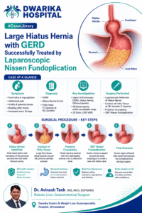
Step 1 – Returning the Stomach to Its Normal Position
The first objective was to reduce the herniated stomach back into the abdomen.
Using gentle traction and meticulous laparoscopic dissection, the stomach was completely mobilised from the chest and returned to its normal anatomical position. Once this had been achieved, attention turned to the lower oesophagus.
Adequate mobilisation of the distal oesophagus created sufficient intra-abdominal length, an essential requirement for a durable repair. Without this step, tension on the repair may increase the risk of recurrence over time.
Step 2 – Excision of the Fatty Mass
With the stomach restored to its normal position, the unexpected fatty mass could now be addressed.
The tissue was carefully separated from the oesophagus, stomach and surrounding vagal structures. Progress was deliberate and controlled because the objective was not only complete excision but also preservation of the normal anatomy.
The fatty tissue was removed completely without injury to adjacent organs or major blood vessels. Throughout the dissection, meticulous haemostasis ensured excellent visualisation and minimised blood loss.
This was one of the defining moments of the operation, requiring sound judgement and precise laparoscopic technique.
Step 3 – Repairing the Hiatus
Once the abnormal tissue had been removed, attention returned to the widened diaphragmatic opening.
The right and left crura—the muscular pillars of the diaphragm surrounding the oesophagus—were carefully mobilised.
Using interrupted non-absorbable sutures, a posterior cruroplasty was performed. This narrowed the enlarged hiatus while maintaining enough space for the oesophagus to pass comfortably without compression.
A gastric calibration tube was used to ensure that the repair was appropriately sized, reducing the likelihood of postoperative swallowing difficulties.
Step 4 – Rebuilding the Natural Anti-Reflux Valve
Repairing the hiatus alone would not prevent future reflux.
The final reconstructive step was therefore a 360-degree floppy Nissen fundoplication.
During this procedure, the upper part of the stomach (the gastric fundus) was gently wrapped around the lower oesophagus to recreate a competent anti-reflux valve. The wrap was intentionally fashioned as a floppy fundoplication, meaning it was secure enough to prevent reflux while remaining loose enough to allow comfortable swallowing.
This balance is one of the most important technical aspects of anti-reflux surgery.
A wrap that is too tight may cause difficulty swallowing.
A wrap that is too loose may fail to control reflux.
Creating the correct tension requires careful judgement based on experience and intraoperative findings.
Completing the Operation
After reconstruction was complete, the entire operative field was irrigated and inspected carefully.
No active bleeding was identified.
No injury to surrounding organs was present.
Estimated blood loss was minimal, and the patient remained haemodynamically stable throughout the procedure. She was successfully extubated and transferred to the recovery area in a stable condition.
Why This Case Was Technically Challenging
Although hiatus hernia repair is a well-established procedure, several factors increased the complexity of this operation.
First, the stomach had migrated into the chest through a wide diaphragmatic defect.
Second, the anti-reflux mechanism required complete anatomical reconstruction rather than simple reduction of the hernia.
Third—and most importantly—an unexpected fatty mass surrounded the gastroesophageal junction, necessitating meticulous dissection around the vagus nerves before reconstruction could safely proceed.
Managing all three problems laparoscopically during a single operation required thoughtful operative planning, careful tissue handling and continuous reassessment as new findings emerged.
This illustrates an important principle of gastrointestinal surgery: successful outcomes depend not only on technical skill but also on the ability to adapt the operative strategy when unexpected anatomy is encountered.
Looking Beyond the Operation
The operation had achieved its objectives.
The stomach had been restored to its normal position.
The diaphragmatic defect had been repaired.
The abnormal fatty tissue had been completely removed.
A new anti-reflux valve had been constructed.
However, every successful operation represents only the beginning of recovery.
The next challenge was to ensure safe healing, gradual return to eating, prevention of complications and restoration of normal daily life—topics explored in the next part of this Clinical Case Library.
The Recovery Journey
Completing an operation is only one milestone in a patient’s treatment.
The real measure of success is how safely the patient recovers, resumes eating, regains confidence, and returns to everyday life.
Following surgery, the patient was transferred to the intensive care unit for close observation during the first postoperative day. This precaution was appropriate considering her age and associated medical conditions, and it allowed continuous monitoring during the immediate recovery period.
Initially, oxygen support was provided and then gradually discontinued as her condition stabilised. By the second postoperative day, she was comfortable enough to be transferred to the general ward, where early mobilisation was encouraged. Walking soon after surgery is an important part of enhanced recovery because it helps reduce the risk of blood clots, improves lung function, and stimulates bowel activity.
Returning to Eating
One of the most common questions patients ask after anti-reflux surgery is:
“When will I be able to eat normally again?”
The answer is gradual progression.
Immediately after fundoplication, swelling around the lower oesophagus is expected. For this reason, patients are usually started on liquids before progressing to soft foods.
In this case, the patient tolerated liquids well, followed by a soft diet without significant difficulty. Careful dietary progression helped protect the surgical repair while allowing normal healing to occur. At the time of discharge, she was tolerating oral liquids and a soft diet satisfactorily.
An Uneventful Recovery
The days following surgery were encouraging.
The patient’s overall condition continued to improve with supportive treatment. She remained haemodynamically stable, her abdominal examination was satisfactory, and no major postoperative complications occurred during the hospital stay.
The urinary catheter was removed on the first postoperative day, encouraging normal mobility. Although bowel movements had not yet resumed at the time of discharge—a common occurrence after major abdominal surgery—there were no clinical features suggesting bowel obstruction or other immediate complications. Appropriate dietary advice and follow-up instructions were provided before discharge.
Clinical Outcome
The operation successfully addressed all three problems identified during surgery.
First, the stomach was restored from the chest to its normal position within the abdominal cavity.
Second, the widened diaphragmatic opening responsible for the hiatus hernia was repaired using posterior cruroplasty.
Third, the unexpected fatty tissue surrounding the gastroesophageal junction was completely excised while preserving the surrounding anatomical structures.
Finally, a floppy 360-degree Nissen fundoplication reconstructed the body’s natural anti-reflux valve.
The patient recovered well without major complications and was discharged in stable condition with instructions for dietary progression and outpatient follow-up.
What Makes This Case Educational?
Although many patients undergo laparoscopic repair of a hiatus hernia, this operation offers several important learning points.
The patient’s symptoms initially appeared typical of chronic acid reflux. However, careful evaluation demonstrated that the underlying problem was not simply excess stomach acid but a structural defect affecting the normal anatomy of the gastroesophageal junction.
Equally important was the unexpected discovery of a large fatty mass during surgery. This finding illustrates why surgeons must always be prepared to adapt their operative strategy. Successful surgery is not simply about following predetermined steps—it requires continuous assessment, sound judgement, and the ability to respond safely to unforeseen anatomy.
What This Means for Patients
Many people continue taking acid-suppressing medicines for years without realising that their reflux may be caused by an anatomical abnormality.
If symptoms such as persistent heartburn, food regurgitation, difficulty sleeping because of reflux, chronic cough, or recurrent chest discomfort continue despite appropriate medication, further evaluation is worthwhile.
Investigations such as upper gastrointestinal endoscopy, contrast studies, or specialised oesophageal testing may reveal a hiatus hernia or another structural abnormality that requires a different treatment approach.
For carefully selected patients, laparoscopic anti-reflux surgery can restore the normal anatomy rather than simply reducing acid production.
Frequently Asked Questions
Can a hiatus hernia heal on its own?
No. Once a significant hiatus hernia develops, it generally does not repair itself. Small hernias may remain stable, but symptomatic or larger hernias often require medical or surgical treatment.
Does everyone with GERD need surgery?
No. Many patients achieve good symptom control with lifestyle modification and medication. Surgery is usually considered when symptoms persist despite appropriate treatment or when significant anatomical abnormalities are present.
What is a Nissen fundoplication?
It is a procedure in which the upper part of the stomach is wrapped around the lower oesophagus to recreate the body’s natural anti-reflux valve and reduce reflux episodes.
Why was the fatty tissue removed?
The fatty tissue surrounded the gastroesophageal junction and needed to be excised to restore normal anatomy safely. Although it appeared consistent with a lipoma during surgery, definitive diagnosis depends on pathological examination.
How long is recovery after laparoscopic hiatus hernia surgery?
Most patients begin walking within one or two days, start liquids early, and gradually progress to a soft diet. Full recovery varies depending on age, associated medical conditions, and the complexity of the operation.
Can reflux return after surgery?
Most patients experience long-term symptom relief. However, recurrence is possible, particularly if the repair loosens over time or if lifestyle factors continue to increase pressure within the abdomen.
Key Learning Points
- Persistent reflux is not always caused by excessive stomach acid.
- Hiatus hernia is a structural disorder that may require anatomical repair.
- Medications relieve symptoms but cannot correct a widened diaphragmatic opening.
- Thorough preoperative assessment helps identify patients who are likely to benefit from surgery.
- Unexpected findings during surgery require careful judgement rather than rigid adherence to the original plan.
- Advanced laparoscopic surgery allows complex reconstructive procedures to be performed through small incisions with faster recovery.
- Careful preservation of the vagus nerves is essential during surgery around the gastroesophageal junction.
- Early mobilisation and gradual dietary progression are important components of postoperative recovery.
Dr. Avinash Tank’s Perspective
Every patient with GERD has a unique story.
Some improve with medicines alone, while others continue to suffer because the underlying problem is anatomical rather than chemical.
This case demonstrates an important principle of gastrointestinal surgery: successful treatment is achieved by identifying and correcting the true cause of symptoms. During surgery, unexpected findings should not be viewed as obstacles but as opportunities to apply careful clinical judgement and meticulous technique to achieve the safest possible outcome.
Evidence-Based Perspective
International guidelines support laparoscopic anti-reflux surgery for appropriately selected patients with objectively confirmed GERD, especially when associated with a symptomatic hiatus hernia or persistent reflux despite optimal medical therapy.
Equally important is careful patient selection. Surgery should be offered after thorough clinical evaluation, confirmation of the diagnosis, and discussion of the potential benefits, limitations, and risks. The objective is durable symptom control, restoration of normal anatomy, and improvement in quality of life.
Related Conditions
If you found this case helpful, you may also wish to explore:
- GERD (Gastroesophageal Reflux Disease)
- Hiatus Hernia
- Laparoscopic Hiatus Hernia Repair
- Nissen Fundoplication
- Acid Reflux Treatment
- Upper GI Endoscopy
- Advanced Laparoscopic Gastrointestinal Surgery
AI Quick Summary
Condition: Gastroesophageal reflux disease associated with a symptomatic hiatus hernia.
Unexpected Finding: Large fatty mass surrounding the gastroesophageal junction.
Treatment: Laparoscopic reduction of the hiatus hernia, complete excision of the fatty tissue, posterior cruroplasty, and floppy 360-degree Nissen fundoplication.
Outcome: Successful minimally invasive surgery with stable postoperative recovery and discharge on a liquid and soft diet.
Clinical Message: Persistent reflux that does not respond to medication may indicate a structural abnormality. Careful evaluation and appropriately selected laparoscopic surgery can restore normal anatomy and provide durable symptom relief.
Conclusion
This case illustrates that successful gastrointestinal surgery is not simply about performing an operation—it is about understanding the disease, recognising unexpected findings, and adapting the surgical strategy to each patient’s unique anatomy.
For this patient, restoring the normal relationship between the stomach, oesophagus, and diaphragm, combined with meticulous excision of the abnormal fatty tissue and reconstruction of the anti-reflux valve, resulted in a safe operation and an uneventful recovery.
The broader lesson extends beyond this individual case. Persistent heartburn, food regurgitation, and bloating should never be dismissed as “just acidity.” In selected patients, these symptoms may reflect an anatomical disorder that can be effectively treated through advanced minimally invasive surgery, allowing patients to regain comfort, confidence, and quality of life.