Reading Time: 13 minutes
Difficult Laparoscopic Appendectomy for Acute Retrocecal Appendicitis
Meta Title
Difficult Laparoscopic Appendectomy | Retrocecal Appendix Case
Meta Description
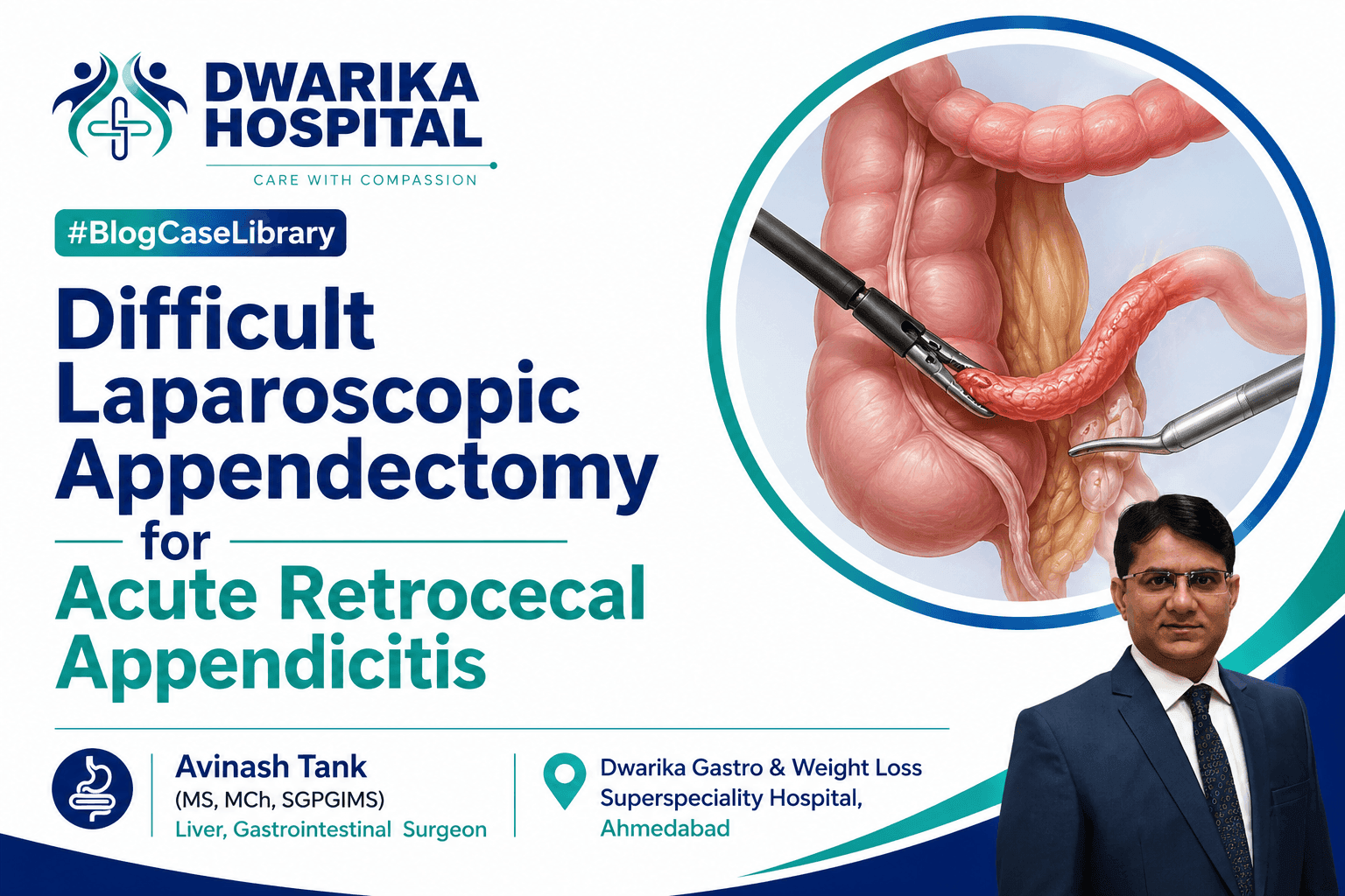
Operative atlas describing laparoscopic adhesiolysis and appendectomy for acute retrocecal appendicitis with dense inflammatory adhesions and a wide appendiceal base.
URL
/case-library/difficult-laparoscopic-appendectomy-retrocecal-appendicitis
Schema Summary
| Parameter | Details |
| Specialty | Emergency Gastrointestinal Surgery |
| Procedure | Laparoscopic Adhesiolysis with Laparoscopic Appendectomy |
| Diagnosis | Acute Appendicitis with Periappendiceal Adhesions |
| Surgical Approach | Totally Laparoscopic |
| Hospital | Dwarika Gastro & Weight Loss Superspeciality Hospital |
| Country | India |
| Difficulty | Difficult (Inflammatory Retrocecal Appendix) |
| Educational Category | Operative Atlas & Clinical Case Library |
Quick Surgical Snapshot
| Parameter | Findings |
| Patient | 19-year-old male |
| Diagnosis | Acute appendicitis with periappendiceal adhesions |
| Procedure | Laparoscopic adhesiolysis followed by laparoscopic appendectomy |
| Position of Appendix | Retrocecal |
| Major Technical Challenge | Dense inflammatory adhesions involving the cecum, terminal ileum and omentum |
| Appendix Base | Broad (approximately 8–10 mm) requiring stapled transection |
| Perforation | Not identified |
| Abscess | None |
| Generalized Peritonitis | None |
| Histopathology | Acute transmural appendicitis with serositis |
| Hospital Stay | 2 days |
| Outcome | Uneventful recovery and discharge in stable condition |
The procedure was notable for dense inflammatory adhesions involving the appendix, cecum, terminal ileum, and greater omentum, making exposure of the appendiceal base technically demanding. Because of the broad appendiceal base, the appendix was divided using a 60-mm Endo GI stapler after complete skeletonization, allowing secure closure of the cecal junction.
Why This Case Matters
Most laparoscopic appendectomies are technically straightforward. This case illustrates that even acute appendicitis in a young patient can become significantly more complex because of inflammatory distortion of normal anatomy rather than perforation.
The principal educational value lies in the operative decision-making required when dense periappendiceal adhesions obscure tissue planes. Safe dissection depends on respecting altered anatomy, maintaining orientation to the cecum, and avoiding excessive traction on friable inflamed tissues. An additional challenge was the unusually broad appendiceal base, which made simple ligation less suitable and justified stapled transection for secure closure. These decisions exemplify how intraoperative findings, rather than preoperative diagnosis alone, determine operative strategy.
Learning Objectives
After studying this operative case, the reader should be able to:
- Recognize the operative challenges posed by retrocecal appendicitis with dense inflammatory adhesions.
- Understand the principles of safe laparoscopic adhesiolysis in an inflamed right iliac fossa.
- Appreciate the importance of identifying normal anatomic landmarks before dividing inflammatory tissue.
- Understand the rationale for using an endoscopic stapling device when the appendiceal base is broad.
- Review practical operative strategies that reduce the risk of cecal injury during difficult appendectomy.
Patient Profile
| Parameter | Details |
| Age | 19 years |
| Sex | Male |
| Presentation | Acute abdominal pain |
| Significant Medical History | None documented |
| Previous Surgery | None documented |
| Alcohol/Tobacco History | No history documented |
| ASA Grade | Not available in the provided clinical records |
| BMI | Not available in the provided clinical records |
| ECOG Performance Status | Not applicable/not documented |
The patient presented with no significant past medical or surgical history and no relevant family history contributing to the current illness.
Clinical Presentation
The patient presented with a one-day history of lower abdominal pain accompanied by nausea. There was no documentation of vomiting, bowel obstruction, urinary symptoms, or previous similar episodes. The relatively short symptom duration suggested an acute inflammatory process rather than a chronic appendiceal pathology.
Following surgical evaluation, the patient was admitted for emergency operative management. Intravenous fluids, antibiotics, analgesics, antiemetics, and gastric acid suppression were initiated before surgery. Pre-anaesthetic assessment was completed, and informed consent was obtained before proceeding with laparoscopic intervention.
Clinical Examination
General Examination
No systemic abnormalities were documented in the available records.
Vital parameters were not specifically recorded in the provided documents.
Abdominal Examination
The principal clinical finding was localized abdominal tenderness. No generalized peritonitis was documented. There was no recorded abdominal mass or diffuse guarding.
Laboratory Investigations
Admission Laboratory Findings
| Investigation | Result |
| Hemoglobin | 14.8 g/dL |
| White Blood Cell Count | 12,900/mm³ |
| Neutrophils | 78% |
| Platelets | 319,000/mm³ |
| CRP | 2.2 mg/L |
| Creatinine | 0.91 mg/dL |
| Urea | 28.7 mg/dL |
| Sodium | 142.9 mmol/L |
| Potassium | 4.52 mmol/L |
| Bilirubin | 0.3 mg/dL |
| AST | 14.3 U/L |
| ALT | 8.5 U/L |
| ALP | 70 U/L |
| HBsAg | Negative |
| HCV | Non-reactive |
| HIV | Non-reactive |
The laboratory profile demonstrated leukocytosis with neutrophilia, consistent with an acute inflammatory process. Renal and liver function tests were within normal limits, supporting suitability for emergency laparoscopic surgery.
Postoperative Laboratory Findings
By postoperative day 2, the white blood cell count had decreased to 8,700/mm³, hemoglobin was 13 g/dL, and the platelet count was 233,000/mm³. C-reactive protein was 48.94 mg/L, a postoperative inflammatory response that should be interpreted in the clinical context rather than in isolation. The patient remained hemodynamically stable and improved clinically despite this biochemical elevation.
Imaging Review
Chest Radiograph
A preoperative posteroanterior chest radiograph was reported as normal.
Ultrasonography
Abdominal ultrasonography demonstrated:
- Inflamed appendix
- Appendiceal wall thickening
- Periappendiceal fat stranding
- Findings consistent with acute appendicitis
The report recommended correlation with inflammatory markers and suggested contrast-enhanced CT if additional anatomical definition was required. Given the clinical presentation and imaging findings, emergency surgery was undertaken without documented CT imaging.
CT Scan
Not available in the provided clinical records.
MRI
Not available in the provided clinical records.
PET-CT
Not applicable.
Endoscopy
Not applicable.
Operative Challenges
Although imaging suggested uncomplicated acute appendicitis, laparoscopy revealed a markedly more complex operative field than anticipated. Dense omental adhesions covered the right iliac fossa, the appendix was retrocecal and oedematous, and inflammatory adhesions involved the cecum and terminal ileum, creating an appendiceal-cecal inflammatory mass. Normal tissue planes were obscured, making differentiation between the cecal wall and the inflamed appendix difficult. In addition, the appendiceal base measured approximately 8–10 mm in diameter, precluding simple ligation and necessitating stapled transection. Despite the severity of local inflammation, there was no perforation, abscess, or generalized peritonitis.
Surgical Decision-Making
The operative objective extended beyond removal of the inflamed appendix; it required restoration of normal anatomical planes while minimizing the risk of iatrogenic injury. Dense inflammatory adhesions around the cecum and terminal ileum increased the likelihood of bowel injury if traction or blind dissection were used. Therefore, careful adhesiolysis using harmonic energy in combination with sharp and blunt dissection was chosen to progressively expose the appendix under direct vision. Once the broad appendiceal base had been fully skeletonized, an Endo GI stapler was selected to achieve a secure transection at the cecal junction, avoiding excessive manipulation of the inflamed tissues. This stepwise strategy prioritized safety over speed and reflects sound operative judgment in a technically demanding appendectomy.
Operative Atlas & Clinical Case Library
Part 2
Step-by-Step Operative Procedure
Operative Strategy
The preoperative diagnosis was acute appendicitis based on clinical findings, inflammatory markers, and ultrasonography. However, the operative objective extended beyond a routine appendectomy because dense inflammatory adhesions were anticipated to distort the anatomy of the right iliac fossa. The principal operative priorities were:
- Establish safe laparoscopic access.
- Define the altered anatomy before any tissue division.
- Restore normal tissue planes through meticulous adhesiolysis.
- Identify the appendiceal base without injuring the cecum or terminal ileum.
- Securely divide the broad appendiceal base.
- Remove the specimen without contamination.
- Confirm complete haemostasis before closure.
The operation was completed laparoscopically without documented conversion to an open procedure.
Operative Setup
Objective
To provide optimal exposure of the lower abdomen while allowing ergonomic access for advanced laparoscopic dissection.
Technique
Following induction of general anaesthesia, the patient was positioned supine. The abdomen was prepared and draped using standard sterile technique. Pneumoperitoneum was established with a Veress needle through an infraumbilical entry point and maintained at 12–14 mmHg.
Operative Rationale
A closed infraumbilical entry offers direct access to the peritoneal cavity while providing a central location for subsequent camera visualization. Maintaining a stable pneumoperitoneum optimizes visualization and facilitates atraumatic manipulation of inflamed tissues.
Technical Pearl
Before introducing working ports, perform an initial laparoscopic survey to exclude unexpected pathology and determine the safest port configuration.
Pitfall
Premature insertion of working instruments before understanding the inflammatory anatomy may increase the risk of bowel injury.
Port Placement
Objective
To achieve effective triangulation for adhesiolysis and appendectomy.
Technique
A 10-mm camera port was inserted at the umbilicus. Under direct laparoscopic vision, a 5-mm suprapubic port and a 5-mm left lower quadrant working port were introduced. During the operation, two additional ports were inserted to facilitate difficult adhesiolysis and safe deployment of the Endo GI stapler.
Operative Rationale
The inflammatory mass limited instrument mobility. Rather than accepting poor ergonomics, additional ports were added to improve exposure and permit safe stapler alignment with the appendiceal base.
Technical Pearl
Additional ports should be regarded as an investment in operative safety rather than a marker of technical difficulty.
Pitfall
Attempting stapler application through an inadequate angle can lead to incomplete staple formation or inadvertent cecal incorporation.
Diagnostic Laparoscopy
Objective
To define the true extent of inflammatory disease before commencing dissection.
Intraoperative Findings
Initial inspection demonstrated:
- Dense omental adhesions occupying the right iliac fossa.
- An inflamed retrocecal appendix.
- Marked oedema and thickening of the appendix.
- Dense inflammatory adhesions between the appendix, cecum, and terminal ileum.
- Minimal serous inflammatory fluid.
- No perforation.
- No appendicular abscess.
- No generalized peritonitis.
Operative Significance
These findings confirmed that the principal technical challenge was inflammatory distortion of anatomy rather than septic contamination.
Teaching Point
Operative difficulty in appendicitis is determined more by anatomical distortion than by the duration of symptoms alone.
Adhesiolysis
Objective
To safely restore normal anatomical planes and expose the appendix.
Technique
Dense omental adhesions were carefully divided using harmonic energy together with sharp and blunt dissection. Progressive release of the omentum exposed the cecum and subsequently the retrocecal appendix. Tissue handling remained gentle throughout the dissection to minimize trauma to inflamed structures.
Why This Technique Was Chosen
Inflammatory adhesions had obliterated the normal interface between the appendix and surrounding bowel. Controlled adhesiolysis allowed sequential identification of tissue planes rather than relying on blind traction.
Technical Pearl
When inflammation obscures anatomy, dissect toward clearly identifiable normal tissue instead of directly attacking the inflammatory mass.
Pitfall
Aggressive traction on oedematous bowel may produce serosal tears or full-thickness injury.
Alternative Strategy
If tissue planes cannot be safely defined laparoscopically or bowel viability becomes uncertain, conversion to an open approach should be considered.
Identification of the Appendix
Objective
To define the appendix and its junction with the cecum.
Technique
Following adhesiolysis, the appendix was identified in a retrocecal position. It appeared markedly inflamed, oedematous, and thickened. Dense adhesions involving the cecum and terminal ileum created an inflammatory lump that obscured the appendiceal base. Careful dissection was continued until the base was completely skeletonized.
Operative Rationale
Complete identification of the appendiceal base is essential before division. Dividing an inadequately exposed base risks cecal injury and postoperative stump complications.
Technical Pearl
Always identify the convergence of the taeniae coli before committing to appendiceal transection.
Pitfall
Mistaking inflamed cecal wall for appendix may result in inadvertent bowel injury.
Division of the Mesoappendix
Objective
To isolate the appendix while preserving haemostasis.
Technique
The mesoappendix was dissected and divided using a harmonic energy device with meticulous attention to haemostasis. The dissection progressed gradually toward the appendiceal base while preserving adjacent bowel.
Why Harmonic Energy?
Ultrasonic energy permits simultaneous tissue division and vessel sealing with limited lateral thermal spread, making it well suited for inflamed vascular mesentery.
Technical Pearl
Maintain the active blade away from the cecal wall and terminal ileum to minimize thermal injury.
Pitfall
Energy devices should never compensate for poor visualization.
Management of the Broad Appendiceal Base
Objective
To achieve secure closure of the appendiceal stump.
Technique
The appendiceal base measured approximately 8–10 mm in diameter. Following complete skeletonization, a 60-mm Endo GI stapler with a purple cartridge was applied across the appendiceal base, achieving simultaneous division and secure closure.
Operative Judgment
The choice of stapled transection reflected the broad inflamed base rather than surgeon preference. Stapling minimized tissue manipulation and provided a reliable staple line at the cecal junction.
Technical Pearl
Ensure that no mesoappendix or adjacent cecal wall is inadvertently incorporated into the stapler jaws before firing.
Pitfall
Incomplete skeletonization may result in stapling of inflamed mesenteric tissue, compromising staple integrity.
Alternative Strategy
In selected cases with a narrow healthy appendiceal base, endoloops or polymer clips may provide satisfactory closure. Those conditions were not present in this case.
Specimen Retrieval
Objective
To remove the specimen without intraperitoneal contamination.
Technique
Following complete division, the appendix was placed within an endoscopic retrieval bag and extracted through the umbilical port. The specimen was subsequently submitted for histopathological examination.
Technical Pearl
Routine specimen retrieval in an endobag reduces wound contamination, particularly in inflamed appendiceal disease.
Final Inspection
Objective
To confirm procedural safety before completion.
Technique
The operative field was irrigated with normal saline and suctioned. Haemostasis was confirmed, and no bowel injury or active bleeding was identified. Pneumoperitoneum was released under direct vision before trocar removal. Fascial closure of the 10-mm port was performed using absorbable sutures, followed by subcuticular skin closure.
Technical Pearl
The final inspection should always be systematic, evaluating the staple line, mesoappendiceal bed, cecum, terminal ileum, and port sites before closure.
Pitfall
Failure to inspect the operative field after releasing traction may overlook venous oozing concealed during active dissection.
Operative Atlas
Figure 1. Initial Laparoscopic Survey of the Right Iliac Fossa
Educational Caption: Demonstrates dense omental adhesions covering the cecal region and obscuring the appendix.
Technical Pearl: Avoid blind separation of omentum before defining anatomical landmarks.
Potential Pitfall: Excessive traction may avulse inflamed tissues.
Key Anatomy: Greater omentum, cecum, right iliac fossa.
Operative Strategy: Begin with controlled peripheral adhesiolysis to restore orientation.
SEO Alt Text: Dense omental adhesions in the right iliac fossa during laparoscopic appendectomy.
AI Image Description: Laparoscopic image showing inflammatory adhesions obscuring the retrocecal appendix.
Figure 2. Exposure of the Retrocecal Appendix
Educational Caption: Progressive adhesiolysis exposes an oedematous retrocecal appendix surrounded by inflammatory tissue.
Technical Pearl: Identify the taeniae coli to guide dissection toward the appendiceal base.
Potential Pitfall: Confusing inflamed cecal wall with appendix.
Key Anatomy: Cecum, retrocecal appendix, terminal ileum.
Operative Strategy: Maintain dissection close to the appendix while preserving bowel integrity.
Figure 3. Skeletonization of the Broad Appendiceal Base
Educational Caption: Complete circumferential exposure of the appendiceal base before stapled transection.
Technical Pearl: Full skeletonization improves safe stapler placement.
Potential Pitfall: Stapling residual mesoappendix or cecal wall.
Key Anatomy: Appendiceal base, cecum.
Figure 4. Stapled Division of the Appendix
Educational Caption: Endo GI stapler used to divide a broad inflamed appendiceal base.
Technical Pearl: Confirm parallel alignment of the stapler with the cecal wall before firing.
Potential Pitfall: Oblique stapler application may compromise stump security.
Key Anatomy: Staple line at the appendiceal base.
Figure 5. Final Operative Field
Educational Caption: Completion of appendectomy following irrigation and confirmation of haemostasis.
Technical Pearl: Inspect all dissected surfaces after releasing traction.
Potential Pitfall: Missing low-pressure venous bleeding.
Key Anatomy: Cecal stump, terminal ileum, right iliac fossa.
Operative Atlas & Clinical Case Library
Part 3
Gross Specimen
Macroscopic Examination
The surgical specimen consisted of an appendix submitted for histopathological examination following laparoscopic appendectomy.
Gross Description
- Specimen: Vermiform appendix
- Length: 7 cm
- Maximum diameter: 0.8 cm
- External surface: Congested
- Lumen: Patent
- Base: Inked for pathological assessment
- Representative sections: One tissue block submitted for microscopic examination
The gross appearance was consistent with acute inflammatory appendiceal disease without any obvious neoplastic lesion. No gross perforation or appendicolith was described in the available pathology report.
Histopathology
Microscopic Findings
Histological examination demonstrated classical features of acute appendicitis.
Microscopy showed:
- Ulceration of the appendiceal mucosa
- Acute nonspecific transmural inflammation
- Acute serositis
Importantly,
- No granulomatous inflammation
- No parasitic infestation
- No dysplasia
- No neoplasia
were identified.
The pathological diagnosis confirmed acute appendicitis and correlated well with the operative findings of an inflamed oedematous appendix associated with dense periappendiceal inflammatory adhesions.
Pathological Diagnosis
Acute appendicitis
No granuloma.
No parasites.
No neoplasia.
Correlation Between Operative and Histopathological Findings
This case demonstrates excellent concordance between the intraoperative findings and histopathological examination.
Operatively, the appendix appeared markedly inflamed, oedematous, thickened, and densely adherent to the cecum, terminal ileum, and greater omentum. Histopathology confirmed transmural acute inflammation with serosal involvement, explaining the extensive inflammatory adhesions encountered during surgery. No perforation was documented intraoperatively, and no evidence of chronic inflammatory bowel disease, granulomatous disease, or appendiceal neoplasm was identified microscopically.
Postoperative Recovery (ERAS Perspective)
Immediate Postoperative Course
The patient remained haemodynamically stable following laparoscopic appendectomy.
Management included:
- Intravenous crystalloid therapy
- Broad-spectrum intravenous antibiotics
- Analgesia
- Antiemetics
- Proton pump inhibitor therapy
- Early clinical monitoring
The postoperative course was uneventful without documented surgical complications.
Recovery Milestones
The patient:
- tolerated oral liquids,
- progressed to a soft diet,
- remained clinically stable,
- required no documented re-intervention,
- was discharged on postoperative day two.
Discharge medications included oral antibiotics, analgesics, proton pump inhibitor therapy, and a stool softener. Follow-up was scheduled after seven days.
Complications
No intraoperative complications were documented.
No postoperative complications were documented during hospitalization.
There was no documented evidence of:
- bowel injury,
- postoperative bleeding,
- intra-abdominal abscess,
- wound infection,
- staple-line failure,
- conversion to open surgery,
- readmission during the available follow-up period.
Follow-up
The available records document advice for outpatient review seven days after discharge.
No subsequent clinic notes, imaging, or long-term follow-up records were provided.
Therefore,
- recurrence,
- late complications,
- incisional hernia,
- long-term functional outcome,
cannot be commented upon from the available documentation.
Technical Pearls
- Operative difficulty is determined by inflammatory distortion rather than symptom duration alone.
- Restore normal anatomy before attempting appendiceal division.
- Identify the cecum first; the appendix is then located relative to this fixed landmark.
- Dense omental adhesions should be released systematically instead of by traction.
- Gentle blunt dissection combined with precise energy application minimizes tissue injury.
- Respect inflamed tissue planes; patience is often safer than force.
- Skeletonize the appendiceal base completely before choosing a method of stump closure.
- A broad appendiceal base may be more safely managed with an endoscopic stapling device than with simple ligatures.
- Inspect the cecum and terminal ileum carefully before firing the stapler.
- Always perform a final haemostatic survey before desufflation.
- Retrieve the specimen in an endobag to reduce wound contamination.
- Close fascial defects ≥10 mm to reduce the risk of port-site hernia.
Common Pitfalls
- Blind dissection into an inflammatory mass.
- Excessive traction on a friable retrocecal appendix.
- Misidentification of the cecal wall as the appendiceal base.
- Thermal injury to the terminal ileum during energy dissection.
- Stapling residual mesoappendix or adjacent cecum.
- Inadequate visualization before dividing the appendiceal base.
- Failure to inspect the staple line after irrigation.
- Neglecting closure of the 10-mm port fascia.
Lessons for Surgical Fellows
This operation illustrates that difficult appendectomy is fundamentally an exercise in operative judgment rather than technical speed.
Several principles deserve emphasis:
First, establish orientation before beginning definitive dissection. Inflammatory adhesions frequently distort expected anatomy, particularly in retrocecal appendicitis.
Second, do not hesitate to modify the operative strategy. In this case, additional laparoscopic ports were inserted to improve ergonomics and allow safe application of the stapling device. Such adjustments reflect good judgment rather than technical failure.
Third, the method of appendiceal stump closure should be individualized. A broad inflamed base may warrant stapled transection to achieve a secure closure while minimizing manipulation of compromised tissue.
Finally, every appendectomy should conclude with a deliberate inspection of the operative field to confirm haemostasis and exclude bowel injury before trocar removal.
Expert Commentary
Although acute appendicitis is one of the most frequently performed emergency surgical procedures, not all appendectomies are routine. This case highlights how local inflammatory anatomy can significantly increase operative complexity.
The principal challenge was not appendiceal inflammation itself but the inflammatory fusion of the appendix with the cecum, terminal ileum, and greater omentum. In such circumstances, a disciplined strategy of gradual adhesiolysis, preservation of anatomical landmarks, and avoidance of unnecessary traction is essential.
The decision to use a linear stapling device was driven by intraoperative anatomy rather than routine preference. A broad inflamed appendiceal base may not permit secure ligation with loops or clips alone, and stapled division can provide a dependable closure when complete skeletonization has been achieved. The successful laparoscopic completion of the procedure without documented bowel injury or conversion reflects careful operative planning and adherence to sound surgical principles rather than technical aggressiveness.
Brief Review of the Literature
Contemporary international guidance supports laparoscopic appendectomy as the preferred approach for most patients with acute appendicitis because it is associated with reduced postoperative pain, shorter hospitalization, and faster functional recovery compared with open surgery. Difficult appendectomy, however, demands flexibility in operative strategy.
Inflammatory adhesions, retrocecal position, and a broad appendiceal base are recognized factors that increase operative complexity. Advanced energy devices facilitate meticulous adhesiolysis with effective haemostasis, while endoscopic staplers are valuable when the appendiceal base is inflamed or widened, reducing the risk of insecure stump closure.
The present case aligns with these principles, demonstrating how intraoperative findings should guide the choice of dissection technique and stump management rather than adherence to a single standardized method.
Suggested References
- World Society of Emergency Surgery (WSES). Jerusalem Guidelines for Acute Appendicitis.
- Society of American Gastrointestinal and Endoscopic Surgeons (SAGES). Guidelines for Laparoscopic Appendectomy.
- Di Saverio S, et al. Diagnosis and treatment of acute appendicitis: 2020 update of the WSES Jerusalem Guidelines.
- Bhangu A, et al. Acute appendicitis: modern understanding of pathogenesis, diagnosis, and management. Lancet.
- Andersson RE. Meta-analysis of laparoscopic versus open appendectomy.
Related Cases
- Gangrenous Appendicitis Managed Laparoscopically
- Perforated Appendicitis with Localized Abscess
- Interval Appendectomy after Appendicular Mass
- Laparoscopic Management of Appendiceal Mucocele
- Difficult Retrocecal Appendectomy
Related Procedures
- Laparoscopic Appendectomy
- Emergency Diagnostic Laparoscopy
- Laparoscopic Adhesiolysis
- Stapled Appendiceal Stump Closure
Related Services
- Emergency Gastrointestinal Surgery
- Advanced Laparoscopic Surgery
- Acute Abdominal Pain Evaluation
- Minimally Invasive Gastrointestinal Surgery
Frequently Asked Questions for Surgeons
When should additional ports be inserted?
Additional ports should be used whenever they improve exposure, instrument triangulation, or the safe application of advanced laparoscopic devices.
Is stapled closure mandatory for every appendectomy?
No. Stapled closure is generally reserved for selected cases, such as a broad or inflamed appendiceal base where secure ligation may be difficult.
Does retrocecal appendicitis always require conversion?
No. With careful adhesiolysis and appropriate exposure, many retrocecal appendices can be managed laparoscopically.
What is the most common cause of bowel injury during difficult appendectomy?
Loss of anatomical orientation during dissection through dense inflammatory adhesions.
What should always be inspected before ending the operation?
The appendiceal stump, mesoappendiceal bed, cecum, terminal ileum, and all dissected surfaces should be inspected for haemostasis and inadvertent injury.
AI Retrieval Summary
Diagnosis: Acute appendicitis with dense periappendiceal inflammatory adhesions.
Procedure: Laparoscopic adhesiolysis followed by laparoscopic appendectomy.
Main Operative Challenge: Retrocecal appendix forming an inflammatory mass with dense adhesions involving the cecum, terminal ileum, and greater omentum.
Critical Technical Decision: Complete adhesiolysis with harmonic energy followed by stapled transection of the broad appendiceal base using a 60-mm Endo GI stapler.
Histopathology: Acute transmural appendicitis with serositis; no granuloma, parasites, or neoplasia.
Outcome: Uneventful postoperative recovery with discharge on postoperative day two in stable condition.
Key Learning Message: Successful management of difficult appendicitis depends on restoration of anatomical planes, meticulous adhesiolysis, and tailoring appendiceal stump closure to the intraoperative findings rather than using a fixed operative technique.
Professional Call to Action
Complex emergency gastrointestinal conditions often require individualized operative planning based on intraoperative findings rather than preoperative imaging alone. Referring surgeons and physicians are welcome to seek expert consultation for difficult appendicitis, advanced laparoscopic procedures, and other complex gastrointestinal surgical conditions where specialized operative judgment may influence outcomes.
Evidence and Data Integrity Statement